Anatomy of the Heart Generator
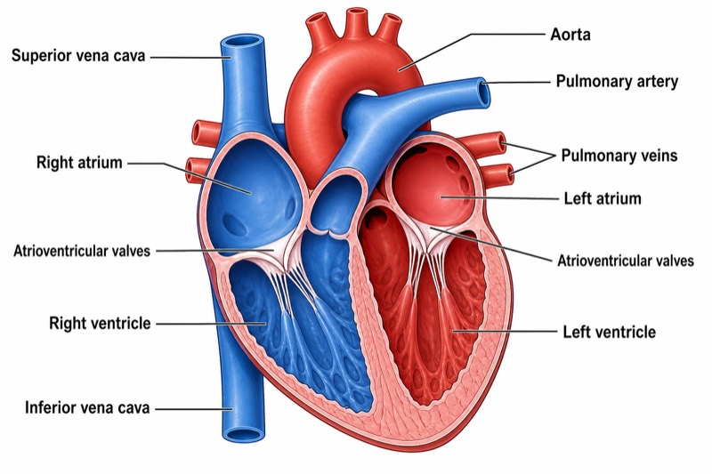
Free online Anatomy of the Heart generator: get a fully labeled figure in about 90 seconds. The AI plans the must-have label list first, then renders a clean textbook-style diagram — every label editable afterwards, ready for papers, assignments and slides.
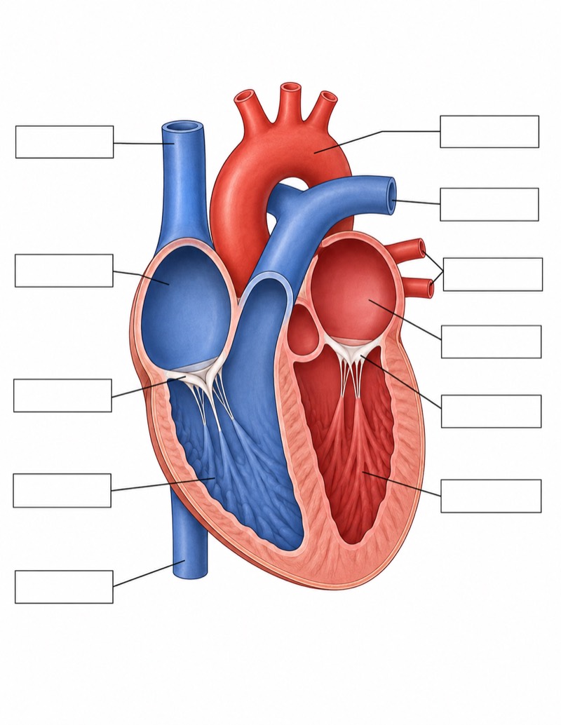
Labels included in this diagram
- Left atrium
- Left ventricle
- Right atrium
- Right ventricle
- Aorta
- Pulmonary artery
- Pulmonary veins
- Superior vena cava
- Inferior vena cava
- Atrioventricular valves
✓ Accurate labels ✓ Edit text after generation ✓ PNG for papers, posters & slides
 OUTPUT · 16:9 · PNG
What this diagram shows
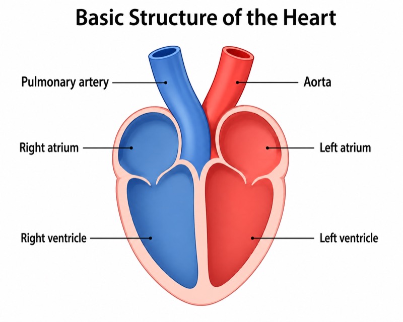
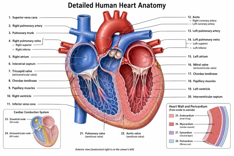
An Anatomy of the Heart diagram shows the four cardiac chambers and the major vessels that connect the pulmonary and systemic circulations. The right atrium receives deoxygenated blood from the superior vena cava and inferior vena cava, while the right ventricle pumps it into the pulmonary artery. Oxygenated blood returns through the pulmonary veins to the left atrium, then enters the left ventricle and is ejected through the aorta. A clear university-level drawing should also indicate the separation of the right and left sides and the relative thickness of the ventricular walls.
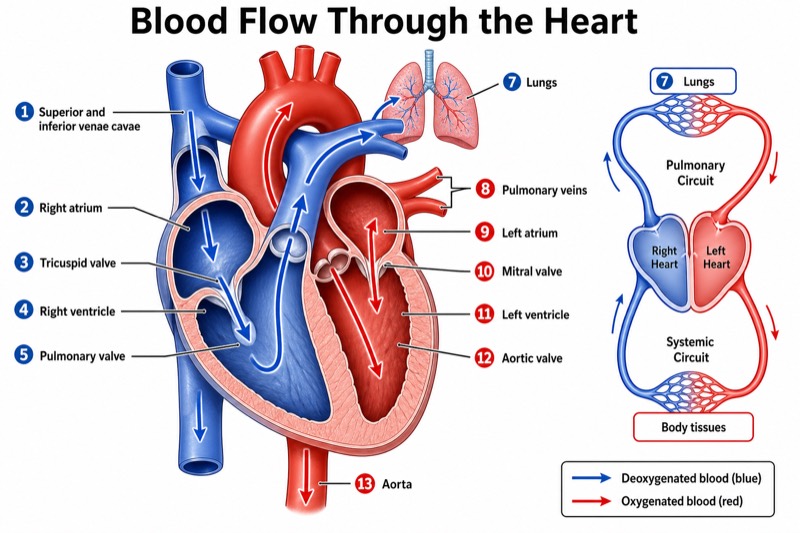
Blood follows a one-way sequence determined by pressure gradients and cardiac valves. It moves from the venae cavae to the right atrium, crosses the right atrioventricular valve into the right ventricle, and exits through the pulmonary artery toward the lungs. Pulmonary veins return oxygenated blood to the left atrium; blood then crosses the left atrioventricular valve into the left ventricle before entering the aorta. The atrioventricular valves prevent ventricular contraction from forcing blood back into the atria. This pathway links pulmonary circulation, where gas exchange occurs, with systemic circulation, which supplies the body.
What a correct diagram must include
- Four chambers — Include the right atrium, right ventricle, left atrium, and left ventricle, arranging each atrium superior to its corresponding ventricle.
- Aorta — Draw it leaving the left ventricle and forming an arch that distributes oxygenated blood to the systemic circulation.
- Pulmonary artery — Show it leaving the right ventricle and dividing toward both lungs; it carries deoxygenated blood despite being an artery.
- Pulmonary veins — Draw vessels returning from the lungs to the left atrium; they carry oxygenated blood despite being veins.
- Venae cavae — Connect the superior vena cava and inferior vena cava to the right atrium to represent venous return from the upper and lower body.
- Atrioventricular valves — Place the tricuspid valve between the right atrium and ventricle and the mitral valve between the left atrium and ventricle.
- Directional arrows — Add arrows from atria to ventricles and from ventricles to arteries so the complete one-way blood-flow sequence is unambiguous.
- Wall proportions and separation — Make the left ventricular wall thicker than the right ventricular wall and keep oxygenated and deoxygenated pathways separated by the cardiac septa.
Common mistakes
- Reversing anatomical left and right — In an anterior-view diagram, the heart's left side usually appears on the viewer's right.
- Coloring vessels by vessel type rather than oxygen content — The pulmonary artery carries deoxygenated blood, whereas the pulmonary veins carry oxygenated blood.
- Connecting the venae cavae to the left atrium — Both the superior and inferior vena cava normally empty into the right atrium.
- Placing atrioventricular valves at arterial outlets — These valves lie between atria and ventricles, not between ventricles and the aorta or pulmonary artery.
- Drawing equal ventricular wall thickness — The left ventricular myocardium should be substantially thicker because it generates pressure for systemic circulation.
Teaching tips
Use the diagram after introducing cardiac orientation and before teaching the cardiac cycle. Ask students to trace one erythrocyte from the superior vena cava through the heart, lungs, and aorta, naming each chamber, vessel, and valve encountered. Follow with questions about oxygenation status, pressure differences, and why the left ventricular wall is thicker. The figure supports assessment of anatomical identification, blood-flow sequence, valve function, pulmonary versus systemic circulation, and the distinction between vessel direction and oxygen content.
FAQ about this diagram
Why is the wall of the left ventricle thicker than that of the right ventricle?
The left ventricle must generate enough pressure to drive blood through the high-resistance systemic circulation. The right ventricle pumps only through the lower-pressure pulmonary circulation, so it requires less myocardial thickness.
Why does the pulmonary artery carry deoxygenated blood while pulmonary veins carry oxygenated blood?
Arteries and veins are named according to the direction of blood flow relative to the heart, not oxygen content. The pulmonary artery carries blood away from the right ventricle, whereas pulmonary veins return blood from the lungs to the left atrium.
What is the correct sequence of blood flow through the heart?
Blood flows from the superior and inferior venae cavae to the right atrium, right ventricle, pulmonary artery, lungs, pulmonary veins, left atrium, left ventricle, and aorta. Atrioventricular valves maintain forward flow between each atrium and ventricle.