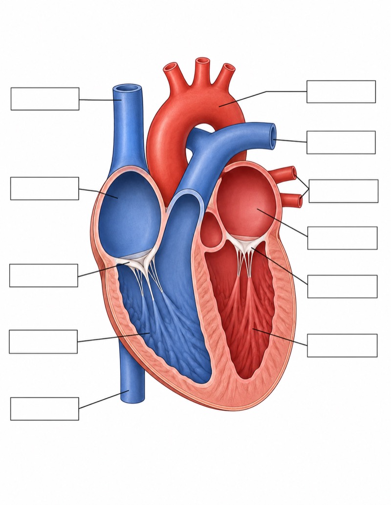
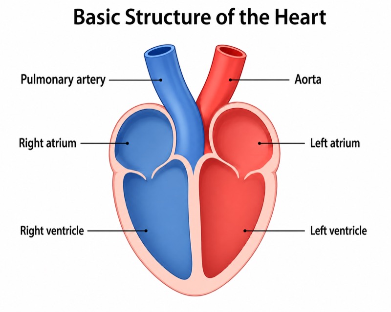
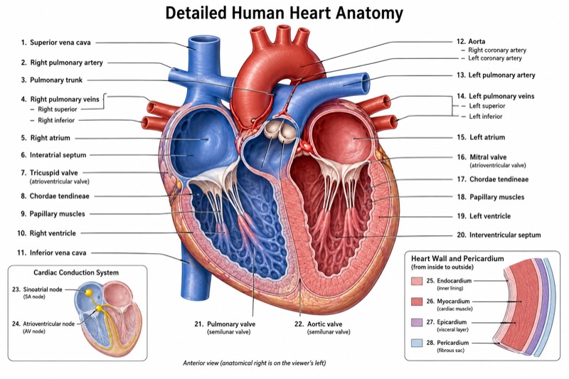
Blood Flow Through the Heart Diagram
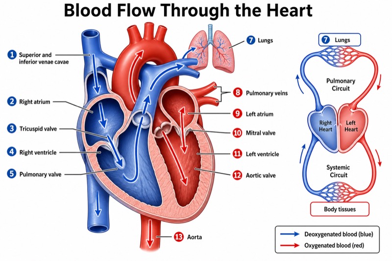
Blood Flow Through the Heart Diagram — an alternative version of the Anatomy of the Heart, generated by AI and fully editable. Download the PNG for quizzes, homework or slides.
Labels included in this diagram
- 1. Superior and inferior venae cavae
- 2. Right atrium
- 3. Tricuspid valve
- 4. Right ventricle
- 5. Pulmonary valve
- 6. Pulmonary arteries
- 7. Lungs
- 8. Pulmonary veins
- 9. Left atrium
- 10. Mitral valve
- 11. Left ventricle
- 12. Aortic valve
- 13. Aorta
- Body tissues
✓ Accurate labels ✓ Edit text after generation ✓ PNG for papers, posters & slides
 OUTPUT · 16:9 · PNG
What this diagram shows
This blood flow through the heart diagram presents a frontal cutaway of the heart with thick, numbered arrows tracing circulation in sequence. Unlike the standard heart anatomy diagram, which emphasizes the positions and structural relationships of chambers, vessels, valves, and tissue layers, this version prioritizes movement and direction. Deoxygenated blood is consistently blue, oxygenated blood is red, and all four valves are labeled with their opening direction. A simplified pulmonary and systemic circulation loop beside the heart provides context beyond the organ itself. Use this version when understanding the route of blood is more important than studying fine anatomical detail.
Read the numbered arrows from the superior and inferior venae cavae to the right atrium, tricuspid valve, right ventricle, pulmonary valve, pulmonary arteries, lungs, pulmonary veins, left atrium, mitral valve, left ventricle, aortic valve, and aorta. Then use the side loop to connect pulmonary circulation with systemic circulation. Compared with the standard main diagram, this pathway-focused version reduces visual competition from secondary anatomy, making sequence errors easier to detect. Trace blue and red routes separately, identify where oxygenation changes, and note that valves maintain one-way flow by opening with forward pressure and closing when pressure reverses.
Teaching tips
- In-class quiz: Remove or cover the numbered labels and ask students to restore the blood-flow sequence, identify each chamber or vessel, and mark where blue blood changes to red.
- Homework assignment: Have students write one sentence for every arrow, including the structure entered, the valve crossed when applicable, and the oxygenation status of the blood.
- Presentation slide: Use this pathway-focused version after the standard anatomy diagram to shift instruction from identifying structures to explaining circulation, pressure-driven valve action, and one-way flow.
- Review outline or printed handout: Pair the diagram with a numbered list of the thirteen major pathway stages so students can rehearse the route aloud and compare pulmonary with systemic circulation.
- Paper or report illustration: Use the simplified red-blue pathway when the text discusses normal cardiac circulation; define the color convention in the caption and avoid treating vessel color as a literal representation of blood appearance.
FAQ about this diagram
How is this version different from a standard heart anatomy diagram?
A standard diagram mainly shows where cardiac structures are located, whereas this version organizes those structures around the ordered movement of blood. Numbered arrows, consistent red-blue coding, valve-opening directions, and the circulation loops make it better for teaching sequence and function.
Why are pulmonary arteries blue and pulmonary veins red in the diagram?
Arteries and veins are classified by flow direction relative to the heart, not by oxygen content. Pulmonary arteries carry deoxygenated blood from the right ventricle to the lungs, while pulmonary veins return oxygenated blood to the left atrium.