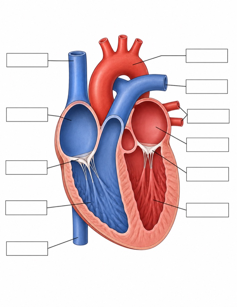
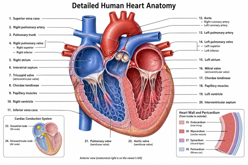
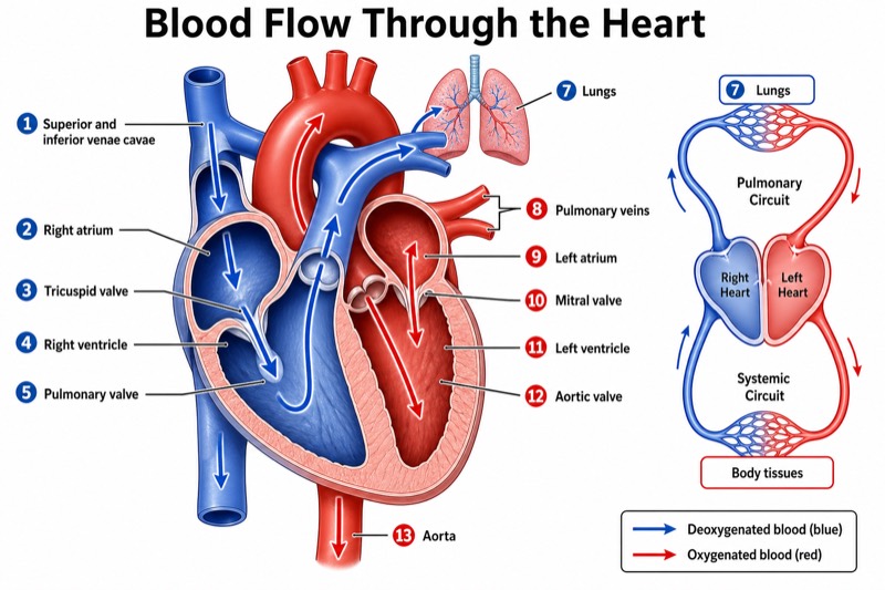
Simple Heart Diagram for Middle School
Simple Heart Diagram for Middle School — an alternative version of the Anatomy of the Heart, generated by AI and fully editable. Download the PNG for quizzes, homework or slides.
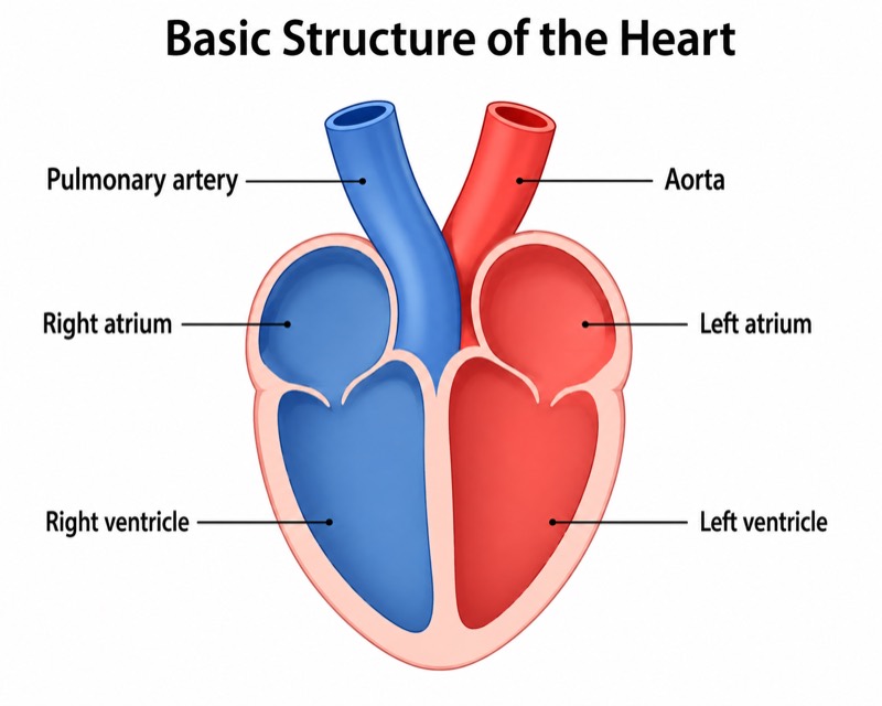
Labels included in this diagram
- Left atrium
- Left ventricle
- Right atrium
- Right ventricle
- Aorta
- Pulmonary artery
✓ Accurate labels ✓ Edit text after generation ✓ PNG for papers, posters & slides
 OUTPUT · 16:9 · PNG
What this diagram shows
This simple heart diagram for middle school shows only six essential structures: the left atrium, left ventricle, right atrium, right ventricle, aorta, and pulmonary artery. Unlike a standard heart anatomy diagram, it omits valves, veins, septal details, coronary vessels, and other features that can distract beginning learners. The frontal cutaway is balanced and spacious, with large labels and red-blue color coding. This reduced version is useful because upper-elementary and middle-school students can first recognize the four chambers and two major arteries before studying more complex anatomy.
Begin by identifying the two atria at the top and the two ventricles below them. Then locate the aorta leaving the left ventricle and the pulmonary artery leaving the right ventricle. Use the red and blue halves as a basic visual guide to the two sides of the heart, while explaining that color represents oxygenation convention rather than the actual color of the chambers. In a frontal view, the heart’s anatomical left usually appears on the viewer’s right. This version supports recognition and orientation; use the standard main diagram when teaching valves, veins, or complete blood-flow pathways.
Teaching tips
- In-class quiz: remove or cover the six labels and ask students to identify the four chambers, aorta, and pulmonary artery within a short time limit.
- Homework: have students color the two sides red and blue, add the six names, and write one sentence explaining which ventricle connects to each artery.
- Presentation slide: display the simplified figure before introducing a standard anatomy diagram, using it to establish chamber position without overwhelming the class with secondary structures.
- Review outline or printed handout: place the diagram beside a six-item vocabulary list so students can match each term to its location and practice anatomical left-right orientation.
- Paper or report illustration: use it for a basic conceptual overview of heart organization, but replace it with a standard detailed diagram when the discussion includes valves, venous return, coronary circulation, or precise blood flow.
FAQ about this diagram
Why does this version omit valves and major veins?
Its purpose is to teach the minimum structural framework: four chambers and two major outgoing arteries. Valves and veins belong in the standard main diagram once students can reliably identify these six structures.
Do the red and blue colors show the heart’s actual appearance?
No. They are conventional teaching colors used to distinguish oxygen-rich and oxygen-poor blood pathways. Students should also remember that anatomical left and right refer to the person shown, not the viewer.