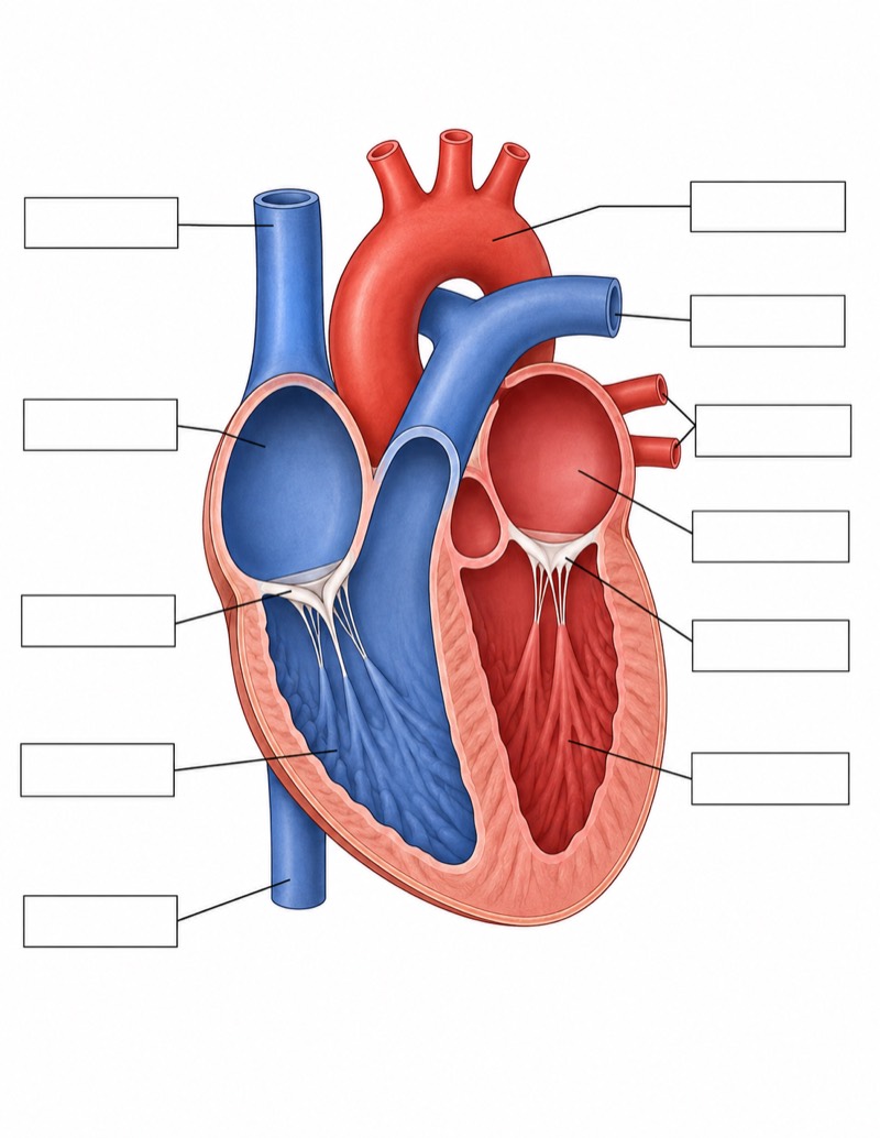
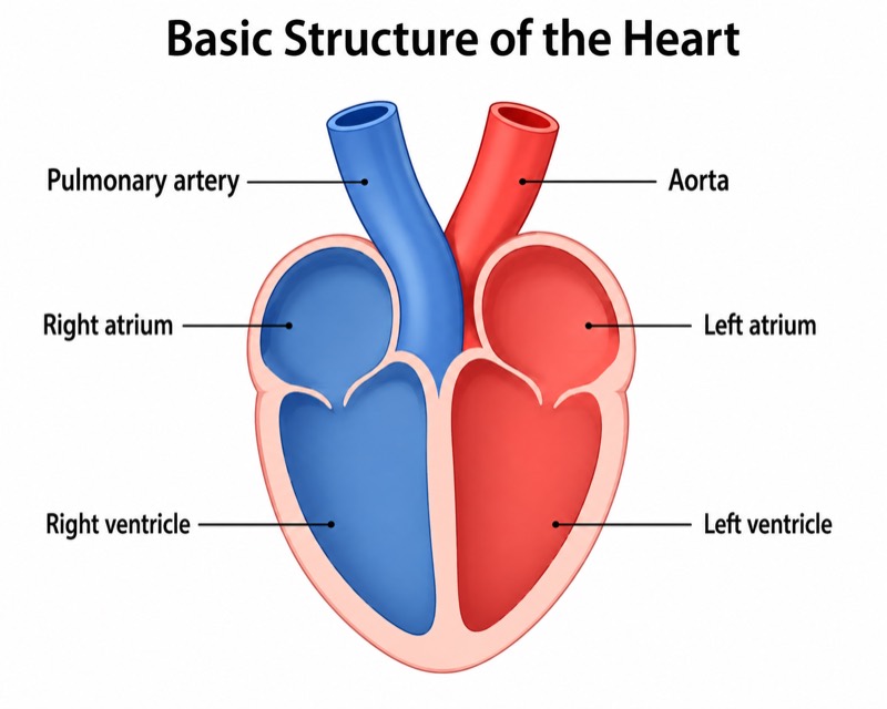
Detailed Heart Diagram (University Level)
Detailed Heart Diagram (University Level) — an alternative version of the Anatomy of the Heart, generated by AI and fully editable. Download the PNG for quizzes, homework or slides.
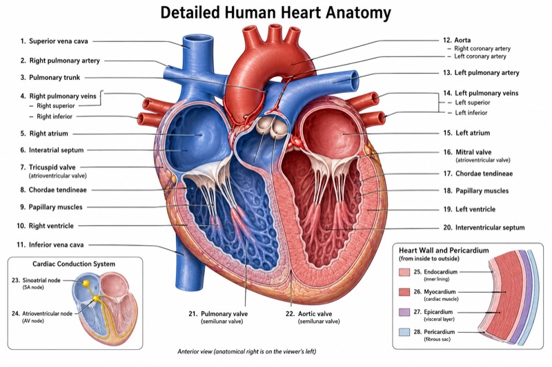
Labels included in this diagram
- Right atrium
- Right ventricle
- Left atrium
- Left ventricle
- Aorta with right and left coronary arteries
- Pulmonary trunk and right and left pulmonary arteries
- Four pulmonary veins: right superior, right inferior, left superior, left inferior
- Superior and inferior venae cavae
- Atrioventricular valves: tricuspid valve and mitral valve
- Semilunar valves: aortic valve and pulmonary valve
- Chordae tendineae and papillary muscles
- Interatrial septum and interventricular septum
- Sinoatrial node and atrioventricular node
- Pericardium and heart wall: endocardium, myocardium, epicardium
✓ Accurate labels ✓ Edit text after generation ✓ PNG for papers, posters & slides
 OUTPUT · 16:9 · PNG
What this diagram shows
This detailed heart diagram (university level) presents the four chambers and major vessels found in the standard overview, but adds the structures needed for advanced anatomical study. It distinguishes the tricuspid, mitral, aortic, and pulmonary valves; chordae tendineae and papillary muscles; interatrial and interventricular septa; four pulmonary veins; superior and inferior venae cavae; right and left coronary arteries; sinoatrial and atrioventricular nodes; and the pericardium. It also identifies the endocardium, myocardium, and epicardium. Unlike the standard main diagram, this version supports structural relationships, tissue-level interpretation, and functional analysis.
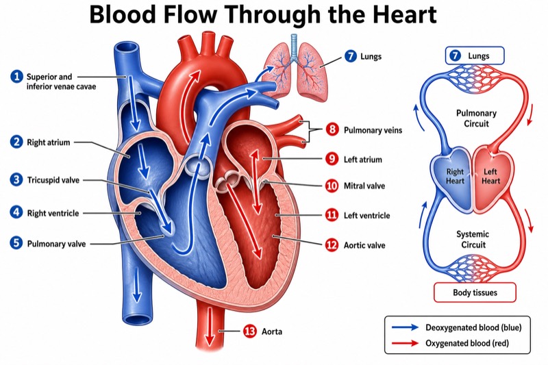
Read the diagram by tracing venous blood from the venae cavae through the right atrium, tricuspid valve, right ventricle, pulmonary valve, and pulmonary artery. Then follow oxygenated blood through the four pulmonary veins, left atrium, mitral valve, left ventricle, aortic valve, and aorta. Use the septa to orient the chambers, and connect the chordae tendineae and papillary muscles with atrioventricular valve control. The conduction nodes indicate where cardiac excitation begins and is delayed. Choose this version instead of the standard main image when precise labeling, flow-path reasoning, or university-level comparison of multiple systems is required.
Teaching tips
- In-class quizzes: remove selected labels and ask students to identify chambers, valves, septa, coronary arteries, conduction nodes, and heart-wall layers rather than testing only the major vessels shown in a standard diagram.
- Homework: have students annotate the complete route of blood flow, using arrows and oxygenation notes while explaining how each valve prevents backflow and how papillary muscles and chordae tendineae stabilize the atrioventricular valves.
- Lecture slides: reveal labels in functional groups—chambers and vessels, valves and supporting structures, conduction tissue, coronary circulation, and wall layers—to build from the standard overview to advanced anatomy.
- Review outlines and printed handouts: pair the figure with a table listing each structure, its location, and its function; use the uncropped layout so noncrossing leader lines remain legible in grayscale printing.
- Research papers or laboratory reports: use the advanced version when the discussion involves cardiac conduction, coronary blood supply, septal anatomy, valve mechanics, pericardial relationships, or the endocardium–myocardium–epicardium organization.
FAQ about this diagram
How is this university-level diagram different from a standard heart anatomy diagram?
A standard diagram usually emphasizes the four chambers, major vessels, and general blood-flow direction. This version adds named valves, valve-support structures, septa, coronary arteries, conduction nodes, pericardium, four pulmonary veins, and the three heart-wall layers, making it more appropriate for university coursework and research-oriented explanation.
Why should I use this detailed version instead of the standard main diagram?
Use the standard main diagram for introductory orientation or a quick explanation of circulation. Use this detailed version when learners must distinguish closely related structures, connect anatomy with valve mechanics and electrical conduction, or prepare accurate labels for assessments, lectures, laboratory work, and academic figures.