Unlabeled Heart Diagram (Blank Worksheet)
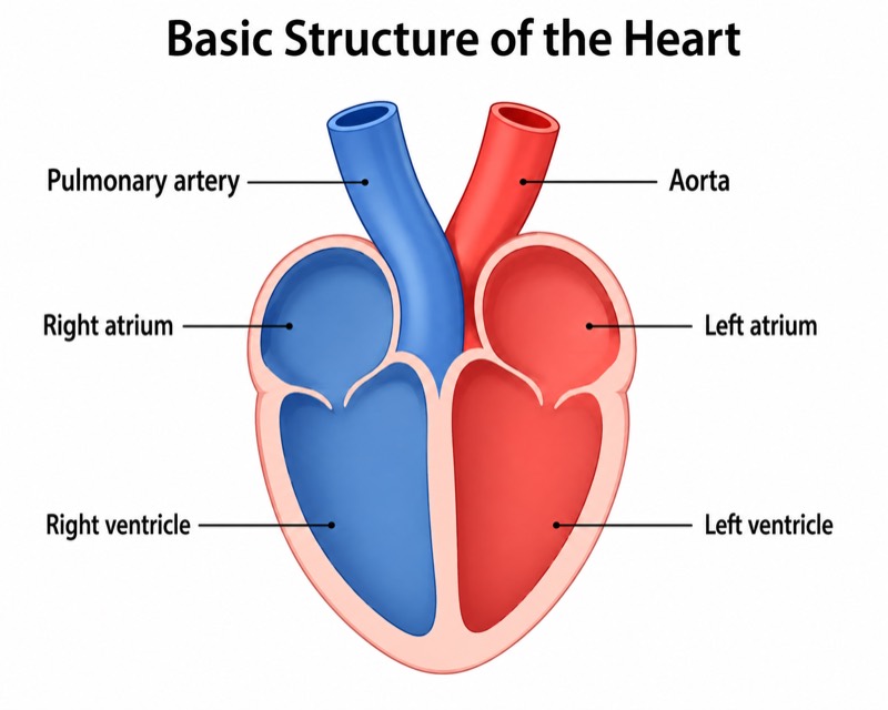
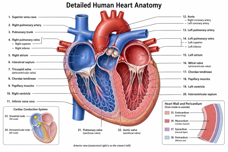
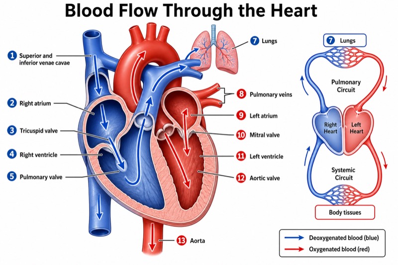
Unlabeled Heart Diagram (Blank Worksheet) — an alternative version of the Anatomy of the Heart, generated by AI and fully editable. Download the PNG for quizzes, homework or slides.
✓ Accurate labels ✓ Edit text after generation ✓ PNG for papers, posters & slides
 OUTPUT · 16:9 · PNG
What this diagram shows
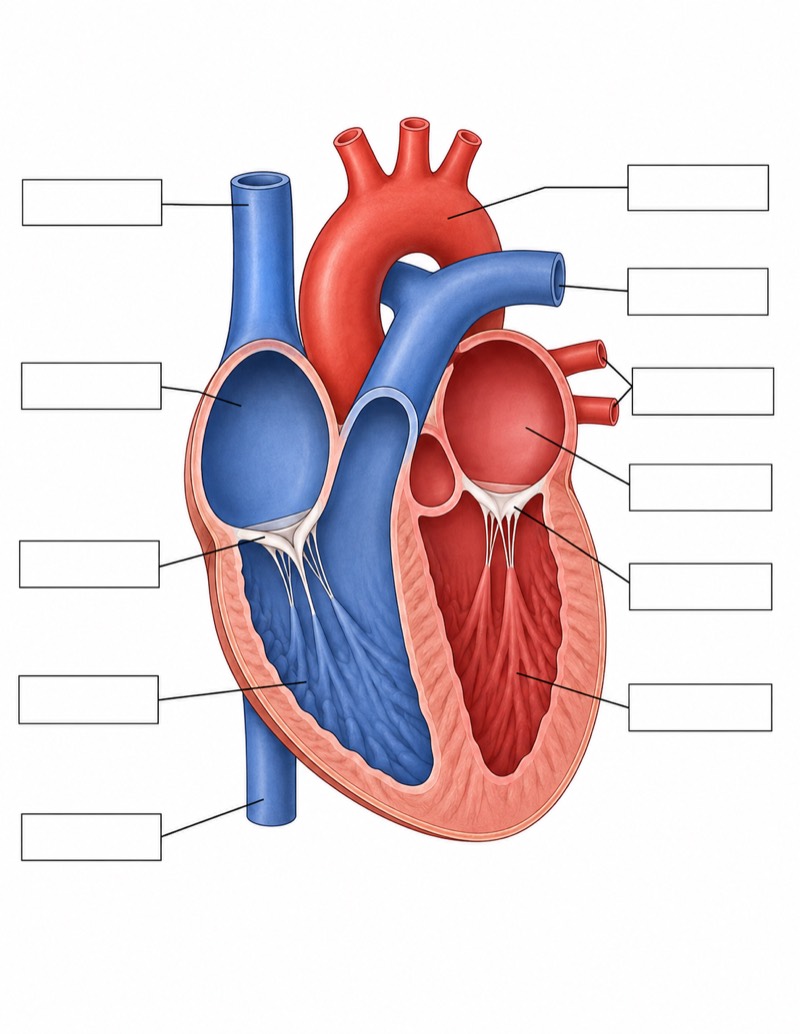
This unlabeled heart diagram (blank worksheet) shows the major chambers, vessels, and atrioventricular valves of the human heart. Its target structures include the left atrium, left ventricle, right atrium, right ventricle, aorta, pulmonary artery, pulmonary veins, superior vena cava, inferior vena cava, and atrioventricular valves. Unlike the standard heart anatomy diagram, it contains no printed names, letters, or numbers. Thin leader lines extend to evenly arranged empty boxes, allowing students to supply every term independently rather than recognize labels that are already visible.
Use the worksheet by tracing each leader line from its blank box to the indicated anatomical structure, then writing the most precise name possible. First identify orientation, remembering that the anatomical right side usually appears on the viewer’s left. Next distinguish the four chambers, follow venous return into the atria, trace ventricular outflow into the pulmonary artery and aorta, and locate the valves between atria and ventricles. This blank version is preferable to the standard labeled figure when recall, self-testing, or formal assessment is the goal, because it removes visual naming cues.
Teaching tips
- Classroom quiz: Print one copy per student and ask learners to complete the blank boxes without notes. Use the standard labeled heart diagram afterward as the answer key and discuss errors involving anatomical right and left.
- Homework assignment: Have students label all chambers, great vessels, and atrioventricular valves, then add arrows showing pulmonary and systemic blood flow. The absence of labels requires active retrieval rather than passive copying.
- Lecture slides: Display the blank worksheet before revealing the standard main diagram. Invite students to identify one structure at a time, using the empty boxes for progressive annotation during instruction.
- Review guide or printed handout: Include the unlabeled figure beside a word bank or a short list of functional clues. Students can repeatedly cover, label, check, and correct the diagram while preparing for an anatomy examination.
- Paper, report, or presentation figure: Use the clean diagram as a base for adding only the structures relevant to a specific discussion, such as ventricular flow or venous return. This avoids the visual crowding of a fully labeled standard figure, but all added annotations should remain anatomically accurate.
FAQ about this diagram
Why use an unlabeled heart worksheet instead of the standard labeled diagram?
The standard diagram supports initial learning by pairing structures with their names. The blank worksheet removes those cues, making it better for retrieval practice, quizzes, and checking whether students can identify anatomy independently.
Which structures should students be able to label on this worksheet?
Students should identify the four chambers, the aorta, pulmonary artery, pulmonary veins, superior vena cava, inferior vena cava, and the atrioventricular valves. In more advanced use, they can also explain blood-flow direction and distinguish oxygenated from deoxygenated pathways.