Human Digestive System Generator
Free online Human Digestive System generator: get a fully labeled figure in about 90 seconds. The AI plans the must-have label list first, then renders a clean textbook-style diagram — every label editable afterwards, ready for papers, assignments and slides.
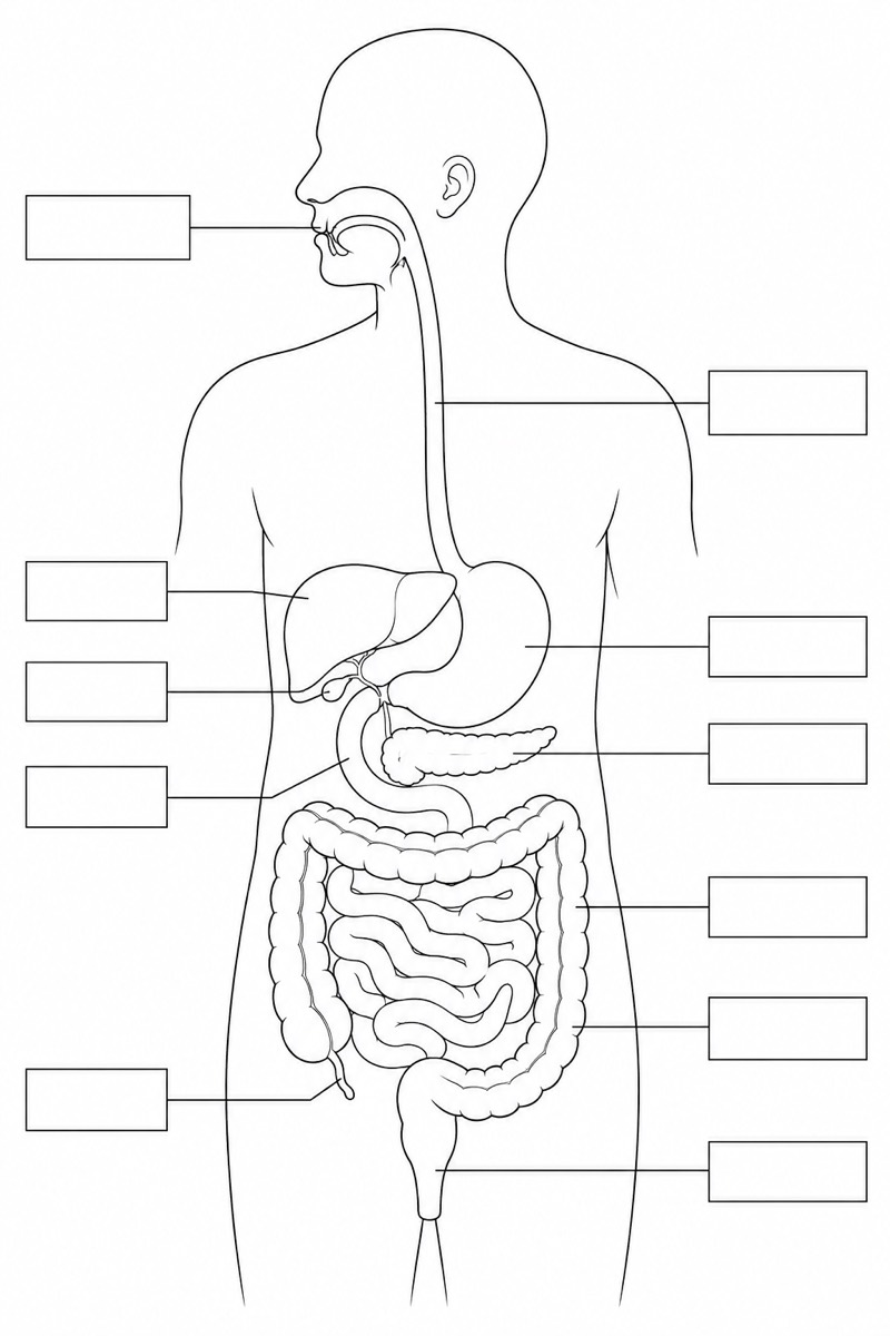
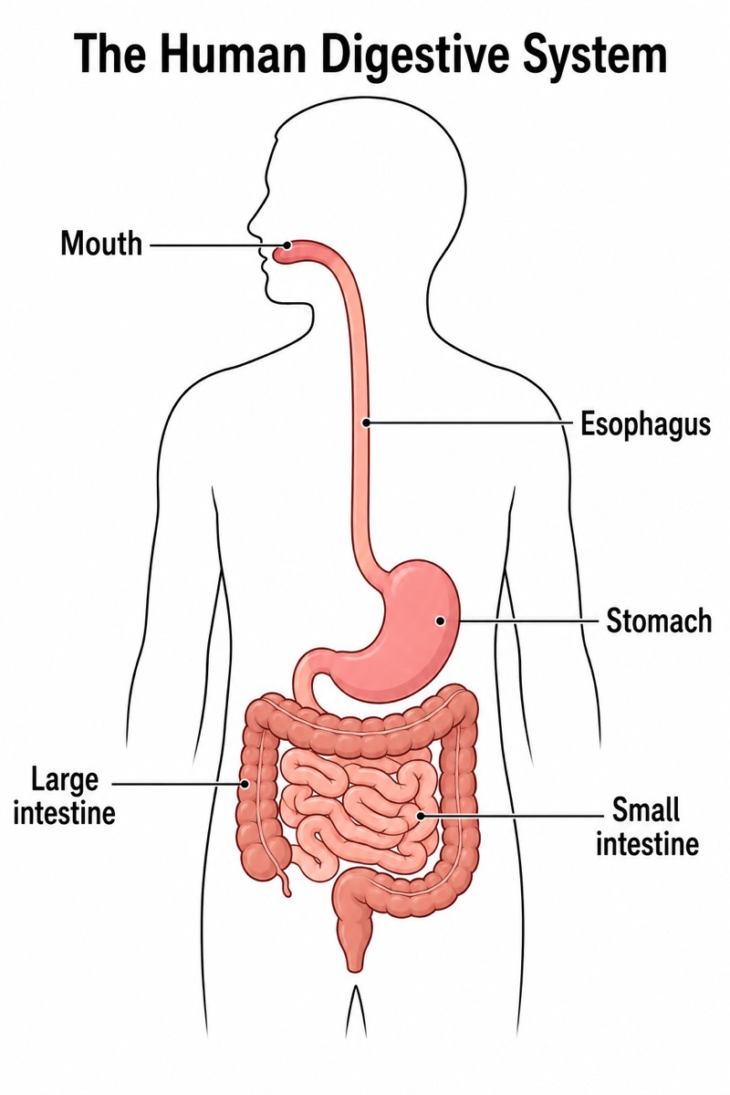
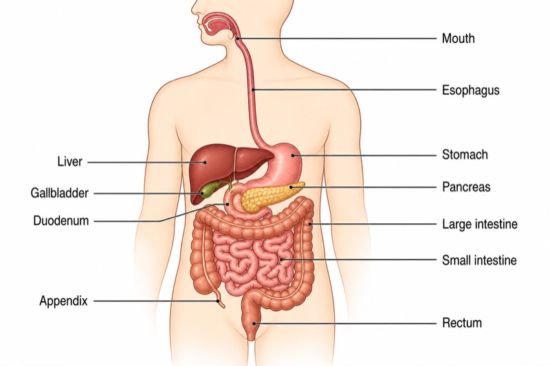
Labels included in this diagram
- Mouth
- Esophagus
- Stomach
- Liver
- Gallbladder
- Pancreas
- Duodenum
- Small intestine
- Large intestine
- Appendix
- Rectum
✓ Accurate labels ✓ Edit text after generation ✓ PNG for papers, posters & slides
 OUTPUT · 16:9 · PNG
What this diagram shows
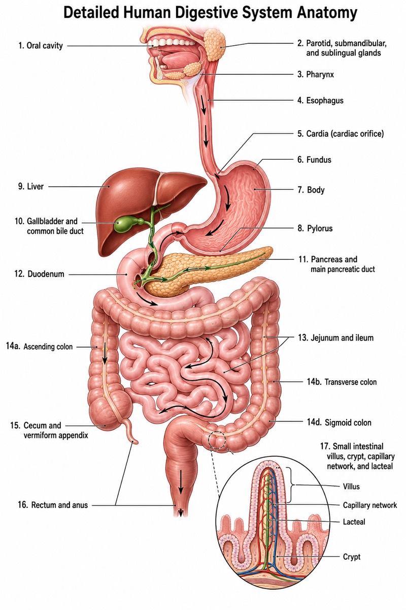
This diagram shows the main organs of the human digestive system and their anatomical positions. Food enters through the mouth, passes down the esophagus, and reaches the stomach. The liver, gallbladder, and pancreas are accessory organs: food does not pass through them, but their secretions help digestion. After the stomach, chyme enters the duodenum, the first part of the small intestine, and then moves through the rest of the small intestine. The large intestine surrounds much of the small intestine. The appendix is a small, narrow tube attached near the beginning of the large intestine, while the rectum stores feces before elimination.
The system works as a continuous pathway with supporting organs. Mechanical digestion begins in the mouth through chewing, while saliva starts the chemical digestion of starch. The stomach churns food and mixes it with gastric juice. In the duodenum, bile from the liver, stored in the gallbladder, helps emulsify fats, and pancreatic juice supplies digestive enzymes. Most digested nutrients are absorbed through the walls of the small intestine into the blood or lymph. The large intestine absorbs water and forms feces, which move into the rectum and leave the body through the anus.
What a correct diagram must include
- Mouth and esophagus: Draw the mouth at the entrance and the esophagus as a narrow tube leading downward to the stomach.
- Stomach: Place it below the esophagus and show its curved sac-like shape, because it stores, mixes, and partially digests food.
- Liver, gallbladder, and pancreas: Place these accessory organs beside or below the stomach, showing that their secretions enter the duodenum rather than food passing through them.
- Duodenum and small intestine: Show the duodenum immediately after the stomach and the small intestine as many coils, because most chemical digestion and nutrient absorption occur there.
- Large intestine: Draw it as a wider outer frame around the small intestine, because it mainly absorbs water and forms feces.
- Appendix: Attach it near the beginning of the large intestine, not to the stomach or small intestine, because it is a small projection from the cecal region.
- Rectum: Place it at the lower end of the large intestine as the storage area for feces before elimination.
- Arrows and labels: Use arrows in the correct sequence from mouth to rectum and keep labels readable without confusing digestive organs with accessory organs.
Common mistakes
- Drawing the esophagus as a branch of the trachea or placing it in front of the airway; the esophagus is the food tube behind the trachea.
- Showing food passing through the liver, gallbladder, or pancreas; these are accessory organs that release helpful substances into the duodenum.
- Putting the large intestine inside the small intestine; anatomically, the large intestine forms a wider outer frame around the coiled small intestine.
- Placing the appendix at the end of the digestive tract; it is attached near the beginning of the large intestine.
- Reversing the sequence of the duodenum and stomach or omitting the duodenum; the duodenum is the first section of the small intestine immediately after the stomach.
Teaching tips
Use the diagram during the introduction to identify structures, then ask students to trace the path of a bite of food with arrows. Ask which organs food actually passes through and which organs provide digestive secretions. Students can match each organ with its main function, such as chewing, mixing, enzyme action, nutrient absorption, or water absorption. The diagram supports common Grade 7 assessment points: digestive tract sequence, the role of accessory organs, and the distinction between digestion and absorption.
FAQ about this diagram
What is the difference between digestion and absorption?
Digestion breaks large food molecules into smaller soluble molecules that the body can use. Absorption is the movement of these digested nutrients through the intestinal wall into the blood or lymph, mainly in the small intestine.
What do the liver, gallbladder, and pancreas do?
The liver produces bile, which helps break large fat droplets into smaller droplets. The gallbladder stores and releases bile, while the pancreas releases digestive enzymes and alkaline fluid into the duodenum.
Why is the small intestine important?
The small intestine completes much of chemical digestion and absorbs most nutrients. Its long, folded structure and many villi provide a large surface area for efficient absorption.