Detailed Digestive System Diagram (University Level)
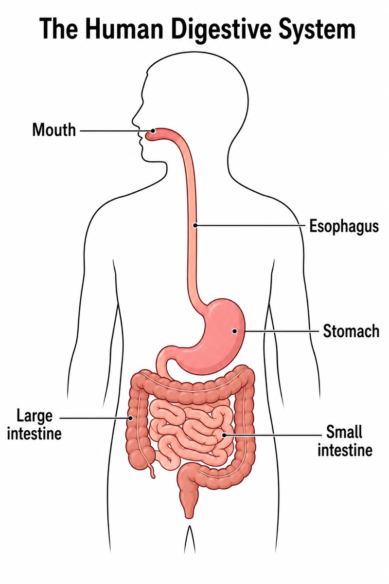
Detailed Digestive System Diagram (University Level) — an alternative version of the Human Digestive System, generated by AI and fully editable. Download the PNG for quizzes, homework or slides.
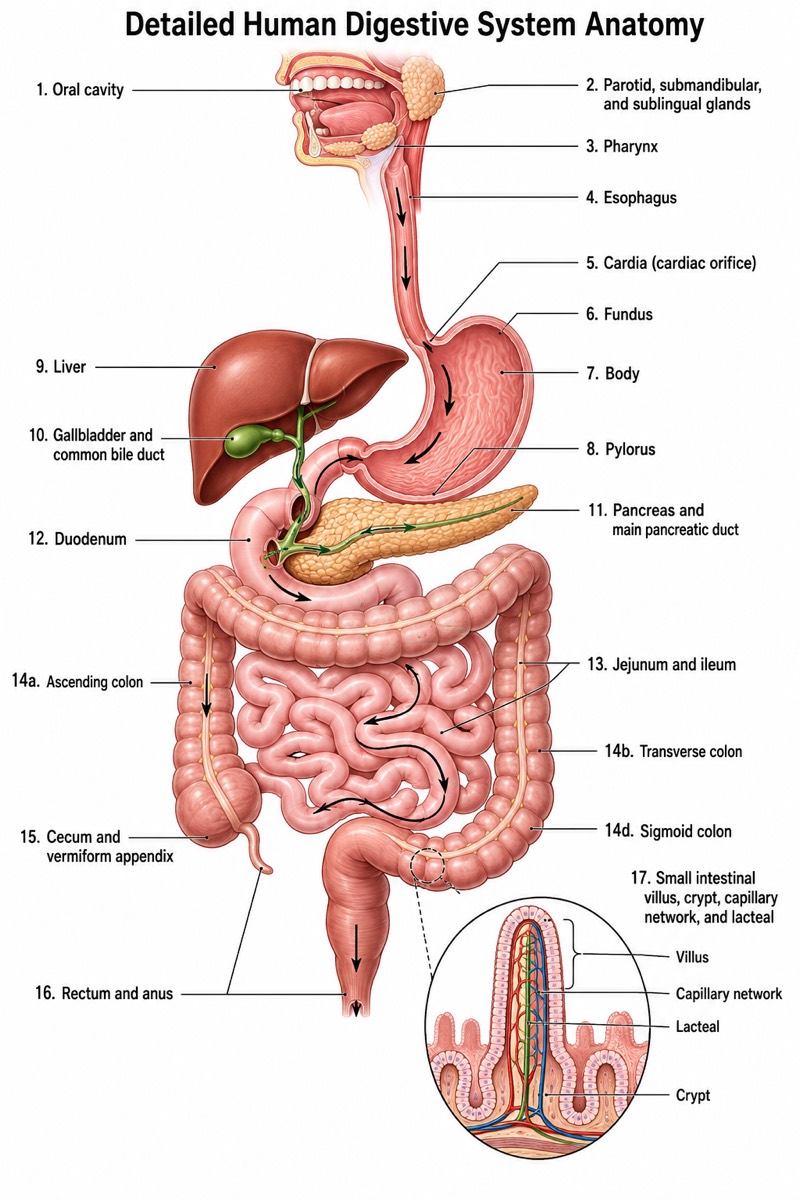
Labels included in this diagram
- Oral cavity
- Parotid, submandibular, and sublingual glands
- Pharynx
- Esophagus
- Cardia, fundus, body, and pylorus of stomach
- Liver
- Gallbladder and common bile duct
- Pancreas and main pancreatic duct
- Duodenum
- Jejunum and ileum
- Cecum and vermiform appendix
- Ascending, transverse, descending, and sigmoid colon
- Rectum and anus
- Small intestinal villus, crypt, capillary network, and lacteal
✓ Accurate labels ✓ Edit text after generation ✓ PNG for papers, posters & slides
 OUTPUT · 16:9 · PNG
What this diagram shows
This detailed digestive system diagram (university level) extends the standard overview by showing more than twelve anatomically organized structures and clinically relevant subdivisions. In addition to the oral cavity, esophagus, stomach, liver, gallbladder, pancreas, intestines, appendix, and rectum, it identifies the parotid, submandibular, and sublingual glands; pharynx; cardia; gastric fundus, body, and pylorus; duodenum, jejunum, and ileum; common bile duct and pancreatic duct; cecum; each colonic segment; anus; and intestinal villi. Use this version when regional anatomy and structural relationships matter more than rapid introductory recognition.
Read the pathway from the oral cavity through the pharynx and esophagus, then follow food through the gastric regions, small-intestinal segments, colon, rectum, and anus. Next, trace accessory secretions from the salivary glands, liver, gallbladder, and pancreas toward the gastrointestinal lumen. The villus inset links organ-level anatomy with absorptive microanatomy. Unlike the standard main diagram, which is best for learning the basic sequence, this version supports comparison of named regions, ducts, junctions, and tissue specializations. Noncrossing leader lines allow each label to be checked without obscuring anatomical position.
Teaching tips
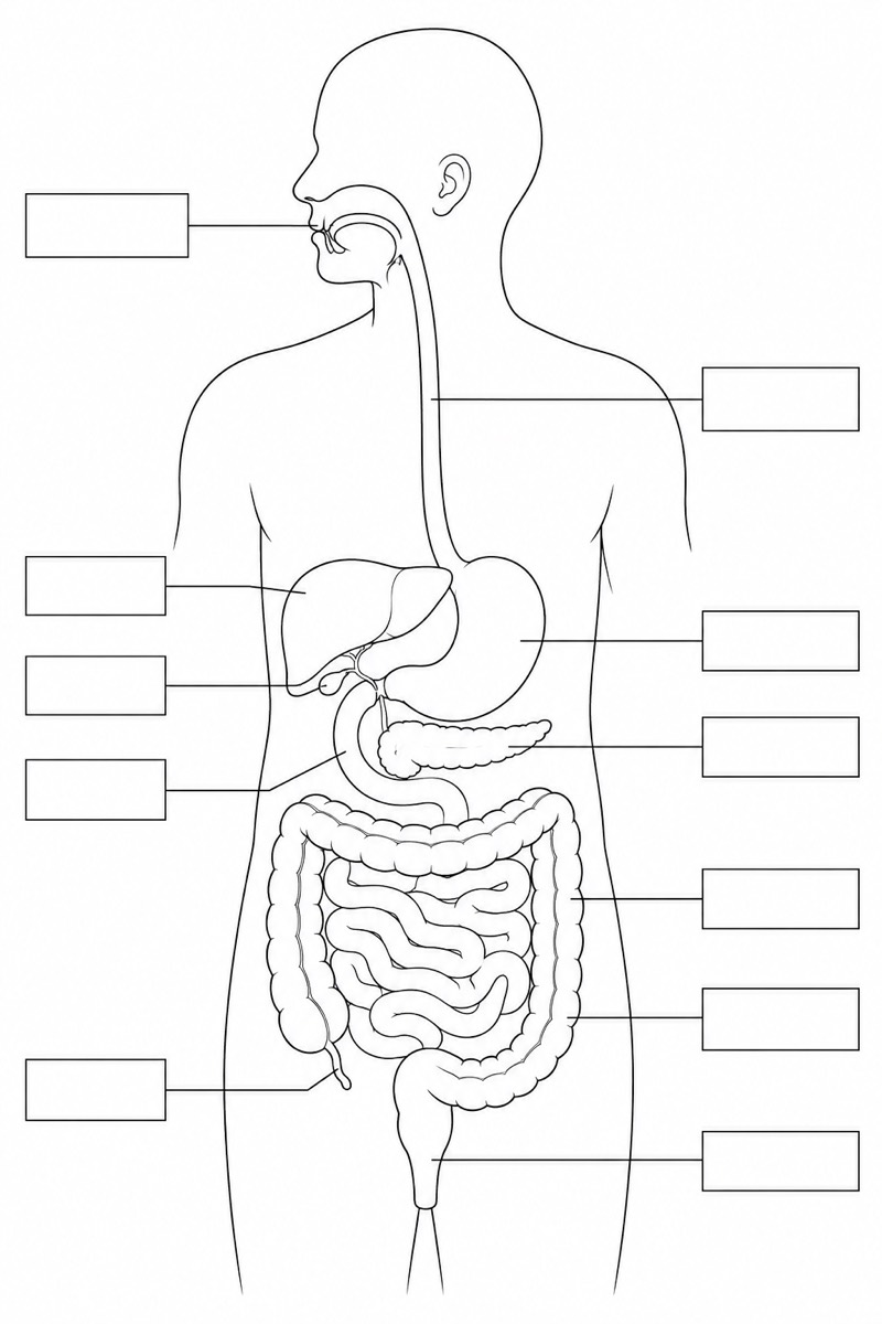
- In-class quizzes: remove selected labels and ask students to identify gastric regions, small-intestinal divisions, colonic segments, salivary glands, and accessory ducts rather than only the major organs shown in the standard diagram.
- Homework: have students annotate the route of a meal in one color and the flow of bile, pancreatic secretion, and saliva in other colors, then explain where chemical digestion and absorption occur.
- Lecture slides: use the full figure after presenting the standard overview, progressively revealing the cardia, pylorus, duodenum, common bile duct, pancreatic duct, and villus inset to connect gross anatomy with physiology.
- Review outlines and printed handouts: pair the labeled image with a blank copy so students can practice spatial recall, distinguish the jejunum from the ileum, and reconstruct the sequence of the large-intestinal segments.
- Research papers and university reports: use the diagram as a general anatomical orientation figure when discussing gastrointestinal physiology, nutrient absorption, hepatobiliary drainage, pancreatic secretion, or regional digestive pathology; cite specialized sources for experimental or disease-specific details.
FAQ about this diagram
Why use this university-level version instead of the standard digestive system diagram?
The standard diagram is better for first learning the major organs and the overall alimentary route. This detailed version is necessary when students must distinguish subdivisions, junctions, glands, and ducts or connect gross anatomy with villus-level absorption.
Which functional relationships should students trace on this diagram?
Students should follow ingested material from the oral cavity to the anus, then separately trace salivary, biliary, and pancreatic secretions into the digestive tract. They should also relate the duodenum to duct entry and the small-intestinal villi to increased absorptive surface area.