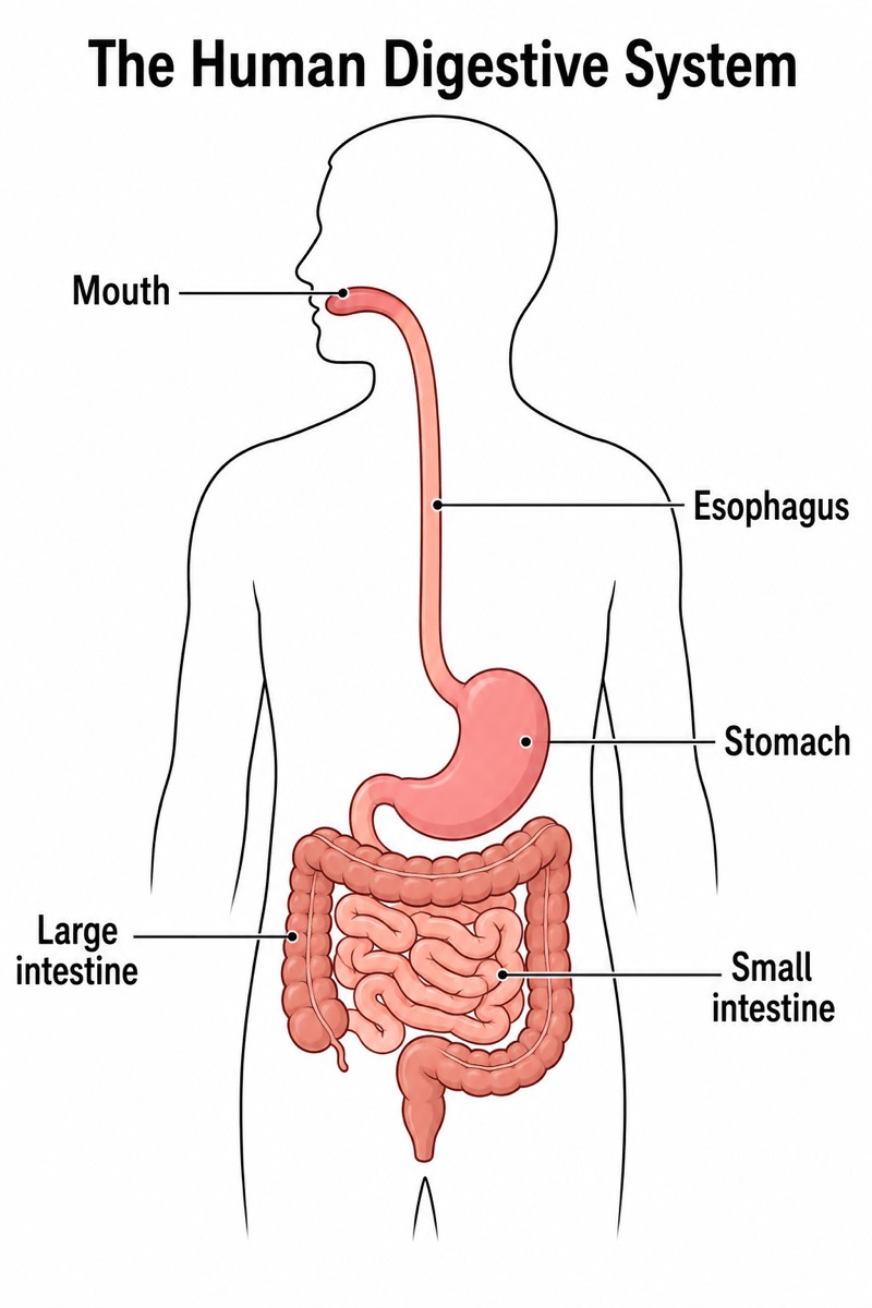
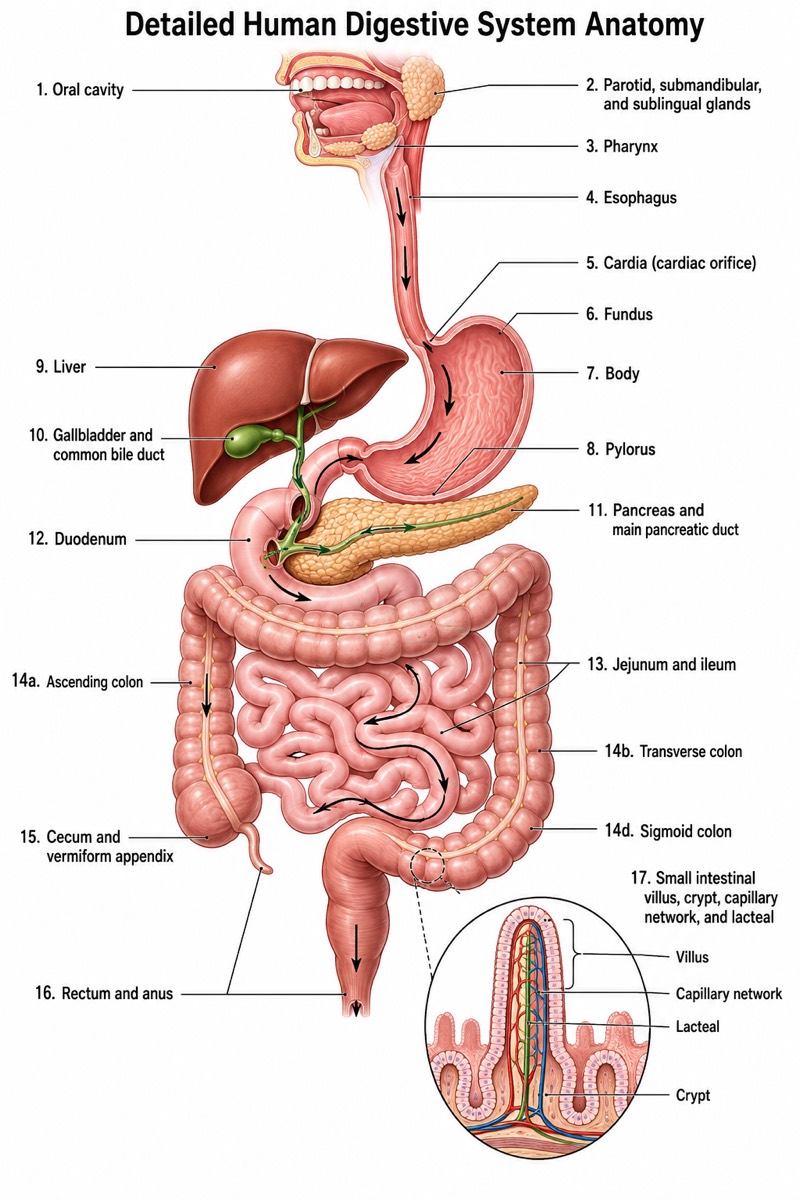
Unlabeled Digestive System Diagram (Blank Worksheet)
Unlabeled Digestive System Diagram (Blank Worksheet) — an alternative version of the Human Digestive System, generated by AI and fully editable. Download the PNG for quizzes, homework or slides.
✓ Accurate labels ✓ Edit text after generation ✓ PNG for papers, posters & slides
 OUTPUT · 16:9 · PNG
What this diagram shows
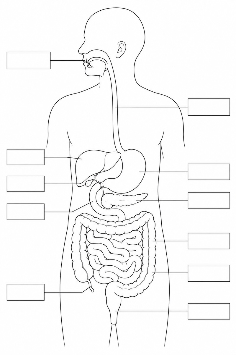
This unlabeled digestive system diagram (blank worksheet) shows the oral cavity, esophagus, stomach, liver, gallbladder, pancreas, small intestine, duodenum, large intestine, appendix, and rectum in their approximate anatomical positions. Unlike the standard main diagram, it contains no printed names, letters, numbers, or answer cues. Each structure is connected to an evenly arranged blank rectangular box by a thin leader line. This version is intentionally incomplete: instead of presenting information for immediate reference, it asks students to retrieve and write the correct anatomical terms.
Read the figure by following each leader line from its blank box to the structure where the line ends. Students can first identify major regions, then distinguish nearby or easily confused organs, such as the stomach and liver, the small and large intestines, or the duodenum and pancreas. Use the blank digestive system worksheet before instruction to assess prior knowledge, during a lesson for guided labeling, or afterward as a retrieval exercise. The standard labeled diagram supports study; this unlabeled version checks whether learners can identify structures without visible prompts.
Teaching tips
- Classroom quiz: Print the diagram and ask students to label all eleven structures from memory within a set time. Grade both anatomical identification and correct spelling.
- Homework assignment: Have students complete the blank boxes, then add arrows showing the path of food from the oral cavity through the esophagus, stomach, intestines, and rectum.
- Presentation or lesson slide: Display the unlabeled figure and reveal organ names verbally or on a separate slide as students identify each structure. This makes the blank version more useful for active participation than the standard labeled main diagram.
- Revision outline: Place the worksheet beside a labeled reference diagram. Students should complete the blank version first, compare their answers, and review errors involving accessory organs, intestinal regions, or anatomical position.
- Printed handout or academic figure: Use the clean, text-free anatomy drawing when a worksheet, assessment appendix, or language-neutral base figure is needed. Add labels separately only when required by the teaching or publication context.
FAQ about this diagram
Why use an unlabeled digestive system diagram instead of the standard labeled version?
A labeled diagram is best for introducing structures and checking terminology, whereas a blank worksheet requires active recall. Removing all names and answer cues makes it possible to assess whether students can independently connect each organ name with its anatomical location.
Which structures should students be expected to label?
The intended answers are oral cavity, esophagus, stomach, liver, gallbladder, pancreas, small intestine, duodenum, large intestine, appendix, and rectum. Teachers may also ask students to classify them as parts of the alimentary canal or as accessory digestive organs.