Structure of a Neuron Generator
Free online Structure of a Neuron generator: get a fully labeled figure in about 90 seconds. The AI plans the must-have label list first, then renders a clean textbook-style diagram — every label editable afterwards, ready for papers, assignments and slides.
Labels included in this diagram
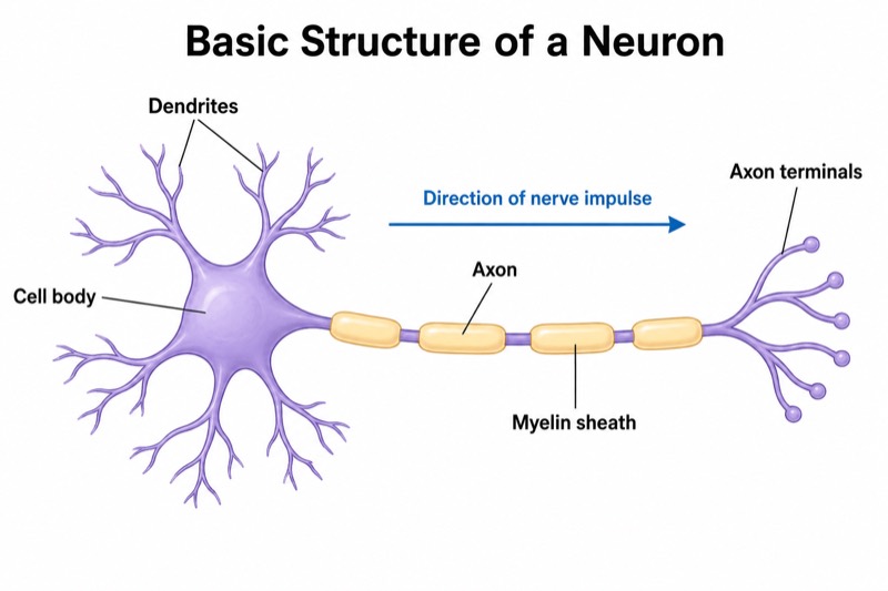
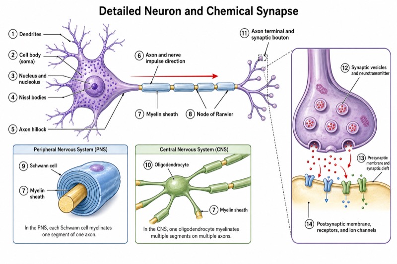
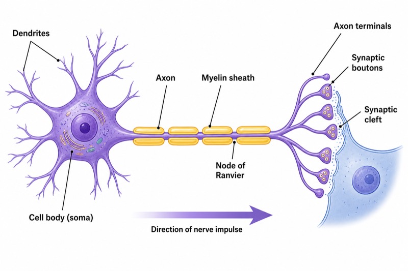
- Cell body (soma)
- Dendrites
- Axon
- Myelin sheath
- Node of Ranvier
- Axon terminals
- Synaptic boutons
- Synaptic cleft
- Direction of nerve impulse
✓ Accurate labels ✓ Edit text after generation ✓ PNG for papers, posters & slides
 OUTPUT · 16:9 · PNG
What this diagram shows
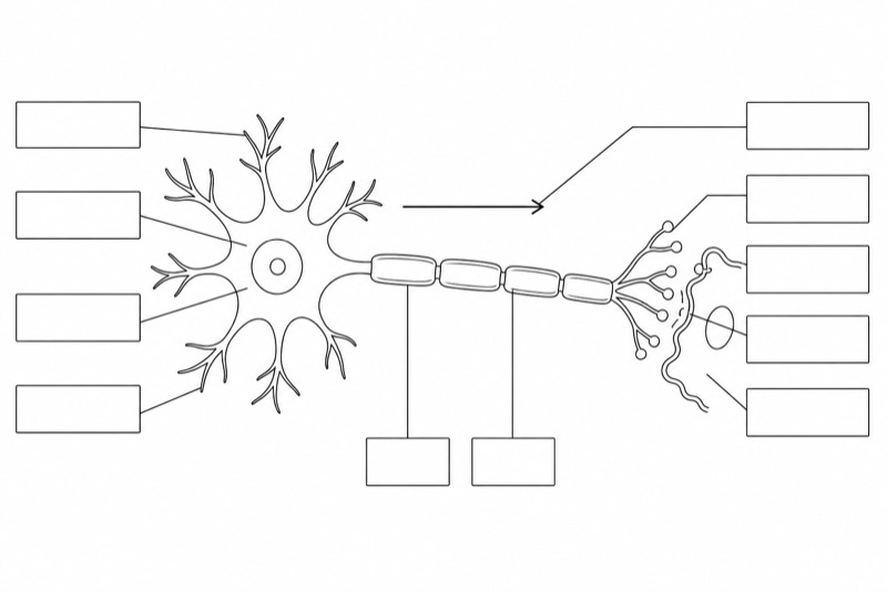
This neuron structure diagram represents the basic organization of a polarized nerve cell and the route by which information is received, conducted, and transmitted. Dendrites extend from the cell body and receive signals from sensory receptors or other neurons. The cell body, or soma, contains the nucleus and integrates incoming information. The axon carries the action potential away from the soma. In a myelinated axon, the myelin sheath electrically insulates the axonal membrane, while Nodes of Ranvier are exposed gaps between myelin segments. At the axon terminals, synaptic boutons release neurotransmitters into the synaptic cleft, allowing communication with the next cell.
The diagram should show a clear functional sequence: dendrites receive input, the cell body integrates graded signals, and the axon conducts an action potential toward the axon terminals. Arrows should point from the dendritic region through the soma and along the axon, ending at the synaptic boutons. Myelin increases conduction efficiency by reducing current loss, and the action potential is regenerated at successive Nodes of Ranvier, producing saltatory conduction. At the terminal, voltage-gated calcium channels trigger synaptic vesicle fusion and neurotransmitter release. Neurotransmitters cross the synaptic cleft and bind receptors on the postsynaptic cell.
What a correct diagram must include
- Cell body (soma): Draw it as the central metabolic and integrative region, and indicate the nucleus if the diagram is sufficiently detailed.
- Dendrites: Show several branching processes arising from the soma because they are the main receptive surfaces for synaptic input.
- Axon: Draw one relatively long process leaving the soma and label it clearly, since it conducts action potentials away from the cell body.
- Myelin sheath: Represent it as segmented insulating layers around the axon rather than as one continuous covering.
- Nodes of Ranvier: Mark the small gaps between adjacent myelin segments because action potentials are regenerated at these exposed regions.
- Axon terminals and synaptic boutons: Place terminal branches at the distal end of the axon and show boutons as specialized swellings that release neurotransmitters.
- Synaptic cleft: Show the narrow extracellular gap between the presynaptic bouton and the postsynaptic membrane, not as a continuation of the axon.
- Impulse direction: Add arrows from dendrites to the soma, along the axon, and toward the axon terminals to represent the usual flow of information.
Common mistakes
- Drawing the axon as a short branch like a dendrite can obscure its conducting role and the usual direction of action-potential propagation.
- Labeling the myelin sheath as part of the Node of Ranvier is incorrect; the node is the unmyelinated gap between neighboring myelin segments.
- Placing the synaptic cleft inside the synaptic bouton is wrong; it is the extracellular space between the presynaptic and postsynaptic membranes.
- Treating dendrites and axon terminals as interchangeable ignores their different roles: dendrites mainly receive input, whereas terminals release neurotransmitter.
- Adding arrows in both directions along one axon without explanation may confuse the normal neuron-to-neuron signaling pathway, which is conventionally shown from soma to axon terminal.
Teaching tips
Use the diagram first to introduce neuronal polarity, then ask students to trace the pathway from dendritic input to neurotransmitter release. Have them distinguish structure from function: which parts receive signals, conduct action potentials, insulate the axon, and communicate with the next cell? The figure supports examination topics including action-potential direction, saltatory conduction, the role of Nodes of Ranvier, calcium-dependent exocytosis, and the difference between a synaptic bouton and the synaptic cleft.
FAQ about this diagram
What is the difference between a synaptic bouton and an axon terminal?
An axon terminal is the distal branching end of an axon. A synaptic bouton is the specialized swelling within that terminal that contains synaptic vesicles, voltage-gated calcium channels, and release machinery.
Why does the impulse appear to jump between Nodes of Ranvier?
The action potential is regenerated at successive exposed nodes, while the myelin sheath limits ion movement across the internodal membrane. This apparent jumping is called saltatory conduction and increases conduction velocity.
Does every neuron have myelin, Nodes of Ranvier, and the same shape?
No. Neurons vary in morphology and may be myelinated or unmyelinated. A typical myelinated neuron diagram is a teaching model that emphasizes rapid axonal conduction rather than representing every neuronal type.