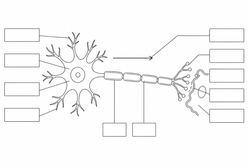
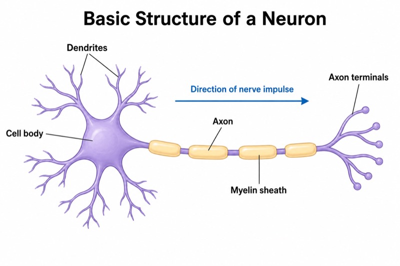
Detailed Neuron Diagram (University Level)
Detailed Neuron Diagram (University Level) — an alternative version of the Structure of a Neuron, generated by AI and fully editable. Download the PNG for quizzes, homework or slides.
Labels included in this diagram
- Cell body (soma)
- Dendrites
- Nucleus and nucleolus
- Nissl bodies
- Axon hillock
- Axon and nerve impulse direction
- Myelin sheath
- Node of Ranvier
- Schwann cell
- Oligodendrocyte
- Axon terminal and synaptic bouton
- Synaptic vesicles and neurotransmitter
- Presynaptic membrane and synaptic cleft
- Postsynaptic membrane, receptors, and ion channels
✓ Accurate labels ✓ Edit text after generation ✓ PNG for papers, posters & slides
 OUTPUT · 16:9 · PNG
What this diagram shows
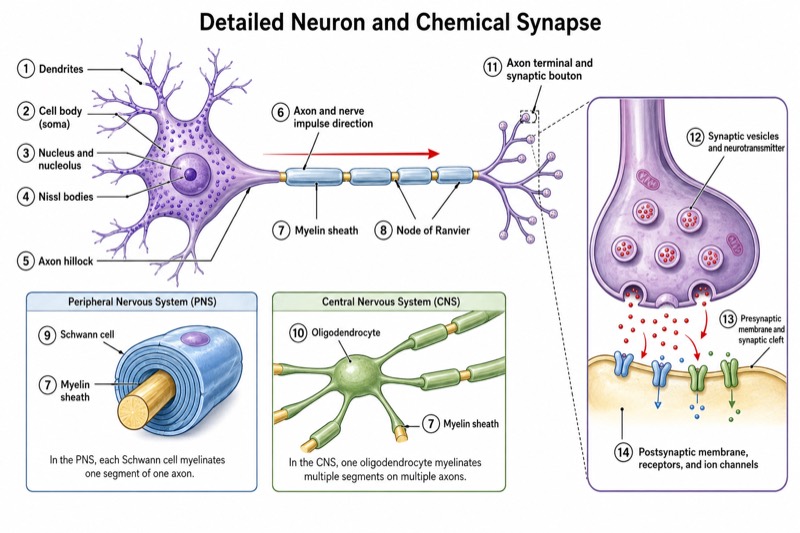
This detailed neuron diagram (university level) extends the standard overview far beyond the basic dendrite–cell body–axon pathway. It labels the soma, dendrites, nucleus, nucleolus, Nissl bodies, axon hillock, axon, myelin sheath, nodes of Ranvier, Schwann cells, oligodendrocytes, axon terminals, and synaptic boutons. An enlarged synapse also identifies synaptic vesicles, neurotransmitters, presynaptic and postsynaptic membranes, the synaptic cleft, receptors, and ion channels. Use this version when learners must connect gross neuronal anatomy with cellular mechanisms rather than simply recognize the major regions of a neuron.
Read the figure from the receptive region toward the output region, following the arrows that show the usual direction of neural impulse transmission: dendrites, soma, axon hillock, axon, and terminals. Then use the synapse enlargement to trace chemical signaling from vesicle fusion and neurotransmitter release to receptor binding and ion-channel activity. Unlike the standard main diagram, this version distinguishes myelinating glia and microscopic synaptic components, making it appropriate for analyzing saltatory conduction, action-potential initiation, and synaptic transmission. The noncrossing leader lines also support accurate labeling practice and rapid comparison among closely related structures.
Teaching tips
- University lecture slides: introduce the standard neuron first, then display this detailed version while explaining action-potential initiation at the axon hillock, saltatory conduction at nodes of Ranvier, and chemical transmission at the synapse.
- In-class quizzes: remove selected labels and ask students to identify at least 12 structures, trace the direction of impulse conduction, and distinguish a Schwann cell from an oligodendrocyte.
- Homework and revision outlines: have students group the labels into receptive, integrative, conductive, myelinating, and secretory components, then add one functional sentence for each group.
- Printed laboratory handouts: use the enlarged synapse for sequencing vesicle docking, neurotransmitter release, diffusion across the synaptic cleft, receptor binding, and postsynaptic ion-channel opening.
- Academic reports or thesis figures: use this version when the discussion requires cellular or synaptic specificity; retain the simpler standard diagram when only overall neuronal polarity or basic anatomy is relevant.
FAQ about this diagram
Why use this university-level diagram instead of a standard neuron diagram?
A standard diagram is best for first learning the major regions, but it usually omits organelles, glial distinctions, and molecular synaptic structures. This detailed version is necessary when studying how anatomy supports action-potential generation, myelination, saltatory conduction, and neurotransmission.
How should Schwann cells and oligodendrocytes be interpreted in the same figure?
Both produce myelin, but Schwann cells myelinate axons in the peripheral nervous system, whereas oligodendrocytes myelinate axons in the central nervous system. Their inclusion makes the diagram comparative; they should not be interpreted as normally myelinating the same axon segment together.