Unlabeled Neuron Diagram (Blank Worksheet)
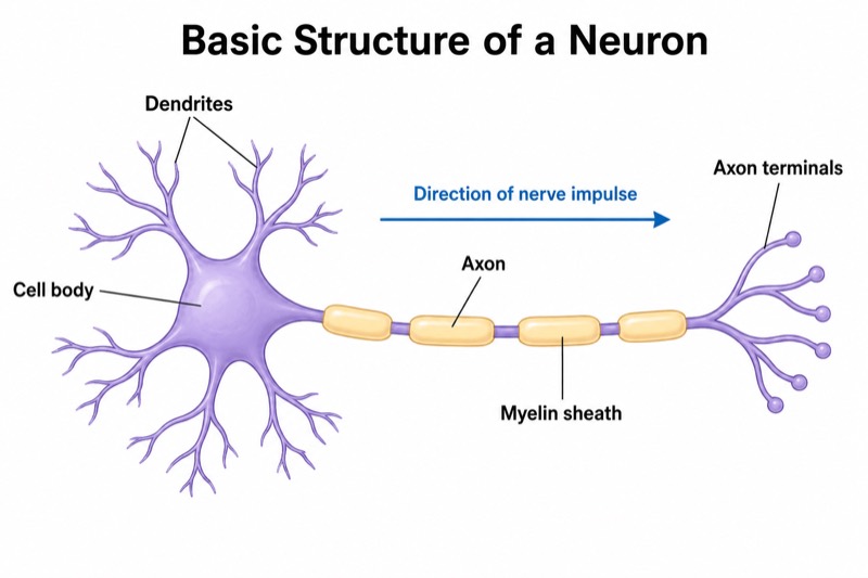
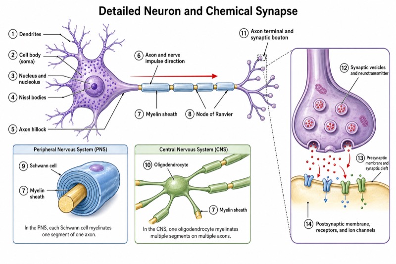
Unlabeled Neuron Diagram (Blank Worksheet) — an alternative version of the Structure of a Neuron, generated by AI and fully editable. Download the PNG for quizzes, homework or slides.
✓ Accurate labels ✓ Edit text after generation ✓ PNG for papers, posters & slides
 OUTPUT · 16:9 · PNG
What this diagram shows
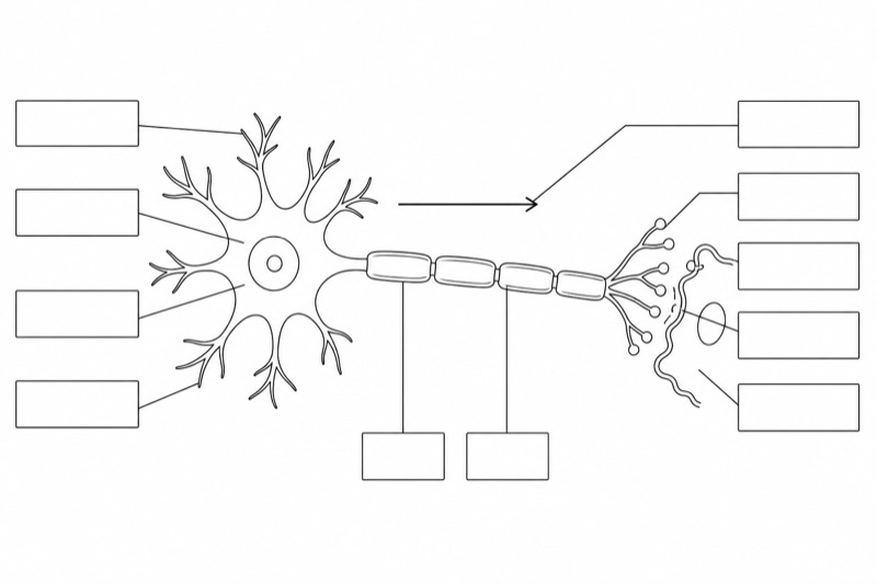
This unlabeled neuron diagram (blank worksheet) shows a neuron with its cell body, dendrites, axon, myelin sheath, nodes of Ranvier, axon terminals, synaptic knobs, and synaptic cleft. Unlike the standard reference diagram, it contains no printed words, letters, or numbers. Thin leader lines connect the structures to evenly arranged empty boxes for handwritten answers. An uncaptioned arrow also indicates the direction of nerve impulse transmission. Removing the completed labels turns the standard teaching image into an active-recall exercise rather than a passive visual reference.
Begin by following the neuron's functional pathway: dendrites receive input, the cell body integrates it, and the axon conducts the impulse toward the terminals. Use shape and position to distinguish myelin segments from the nodes of Ranvier between them, then identify the synaptic knobs and the narrow synaptic cleft. Students should write one anatomical term in each blank box and explain the arrow's meaning separately. This blank version is useful because it tests recognition, spatial relationships, and signal flow without allowing learners to copy labels from the standard main diagram.
Teaching tips
- In-class quiz: distribute the blank worksheet without the standard labeled diagram and ask students to complete every box, then describe the direction of nerve impulse conduction in one sentence.
- Homework assignment: have students label the structures from memory, add a brief function beside each term, and compare their completed sheet with the standard main diagram afterward.
- Presentation slide: display the unlabeled neuron diagram before revealing a labeled reference slide; invite students to identify each structure as a guided retrieval activity.
- Review outline or printed handout: place the blank version next to notes on neuronal signaling so learners can repeatedly practice the anatomy, while reserving the standard diagram for self-checking.
- Paper or laboratory report figure: use the clean blank layout as a base for adding only the structures relevant to the discussion, while defining all added labels and the impulse-direction arrow in the caption.
FAQ about this diagram
Why use a blank neuron worksheet instead of the standard labeled diagram?
The standard diagram supports initial learning by pairing each structure with its name, whereas the blank version requires active recall. It is therefore better for checking whether students can independently recognize structures and reconstruct the pathway of neural transmission.
How should the nerve impulse direction be interpreted when the arrow has no caption?
The arrow should run from the receptive region of the neuron toward its output region: dendrites and cell body, then along the axon to the axon terminals. At the synapse, signaling proceeds from the synaptic knobs across the synaptic cleft toward the next target cell.