The Human Skeletal System Generator
Free online The Human Skeletal System generator: get a fully labeled figure in about 90 seconds. The AI plans the must-have label list first, then renders a clean textbook-style diagram — every label editable afterwards, ready for papers, assignments and slides.
Labels included in this diagram
- Skull
- Clavicle
- Scapula
- Humerus
- Ribs
- Vertebral column
- Pelvis
- Femur
- Tibia
- Fibula
✓ Accurate labels ✓ Edit text after generation ✓ PNG for papers, posters & slides
 OUTPUT · 16:9 · PNG
What this diagram shows
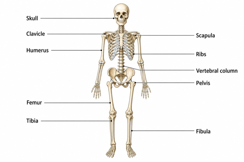
This anterior-view diagram presents the organization of the adult human skeletal system, which typically contains 206 bones. The skull protects the brain and supports the facial framework. The vertebral column forms the central supporting axis, while the ribs enclose much of the thoracic cavity. The clavicle and scapula constitute the pectoral girdle, connecting each upper limb to the axial skeleton. The humerus forms the arm skeleton. Inferiorly, the pelvis transfers body weight to the lower limbs, where the femur forms the thigh and the tibia and fibula form the leg.
The labeled bones illustrate the relationship between the axial and appendicular divisions. The skull, vertebral column, and ribs belong to the axial skeleton, which provides support and protects major organs. The clavicle and scapula connect the humerus to the trunk and permit extensive shoulder movement. The pelvis articulates with the vertebral column and receives the heads of the femora at the hip joints. At the knee, each femur articulates mainly with the tibia. The tibia is the larger, medial, weight-bearing leg bone, whereas the fibula lies laterally and primarily provides muscle attachment and ankle stability.
What a correct diagram must include
- Skull: Draw it at the superior end of the vertebral column and distinguish the cranial vault from the facial skeleton.
- Clavicles and scapulae: Show paired clavicles across the upper chest and place the scapulae posterolaterally, with only part of each visible in an anterior view.
- Vertebral column: Draw a continuous midline axis from the skull to the pelvis, using gentle cervical, thoracic, and lumbar curvatures rather than a straight rod.
- Rib cage: Include bilateral curved ribs attached posteriorly to the thoracic vertebrae and arranged symmetrically around the thorax.
- Humeri: Position one humerus between each shoulder and elbow, with its proximal head directed medially toward the scapular glenoid cavity.
- Pelvis: Represent the pelvic girdle as a strong bilateral ring that supports the trunk and forms the acetabula for the femoral heads.
- Femora: Draw the femur as the long bone of each thigh, angling slightly medially from the hip toward the knee.
- Tibiae and fibulae: Place the larger tibia medially and the slender fibula laterally in each leg, maintaining correct left-right orientation.
Common mistakes
- Placing the fibula on the medial side of the leg; in anatomical position, the tibia is medial and the fibula is lateral.
- Showing the scapula entirely on the front of the thorax; it lies mainly on the posterior thoracic wall and is only partly visible in an anterior diagram.
- Connecting the humerus directly to the clavicle; the humeral head articulates with the glenoid cavity of the scapula.
- Drawing the vertebral column as perfectly straight; its normal sagittal profile includes cervical and lumbar lordoses and thoracic and sacral kyphoses.
- Making the femur vertical from hip to knee; the shaft normally angles medially, helping position the knee beneath the body's center of gravity.
Teaching tips
Use the diagram after introducing anatomical position and directional terminology. Ask students first to identify each labeled bone, then classify it as part of the axial skeleton, pectoral girdle, pelvic girdle, upper limb, or lower limb. Follow with relationship questions such as, “Which bone is lateral in the leg?” and “How is the upper limb attached to the trunk?” The figure supports assessment of bone identification, medial-lateral orientation, skeletal divisions, joint relationships, weight transmission, organ protection, and differences between anterior visibility and actual anatomical position.
FAQ about this diagram
Which labeled bones belong to the axial skeleton?
The skull, vertebral column, and ribs are components of the axial skeleton. They form the central body axis and primarily support posture and protect the brain, spinal cord, and thoracic organs.
What is the functional difference between the tibia and fibula?
The tibia is the larger medial bone and transmits most body weight from the knee to the ankle. The fibula is slender and lateral; it bears little weight but provides muscle attachment and contributes to ankle stability.
How are the upper and lower limbs connected to the axial skeleton?
The upper limb connects through the pectoral girdle, formed by the clavicle and scapula, with the clavicle providing the only direct bony link to the axial skeleton. The lower limb connects through the pelvic girdle, which articulates firmly with the sacrum and transfers body weight to the femur.