Human Respiratory System Generator
Free online Human Respiratory System generator: get a fully labeled figure in about 90 seconds. The AI plans the must-have label list first, then renders a clean textbook-style diagram — every label editable afterwards, ready for papers, assignments and slides.
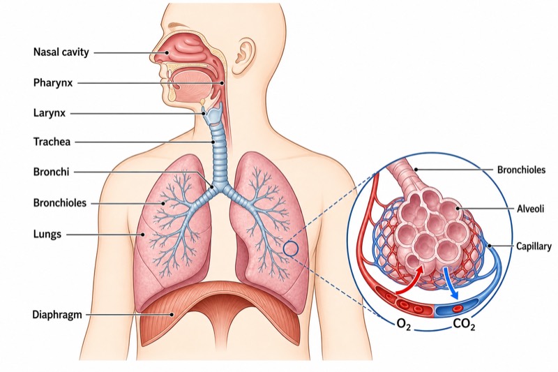
Labels included in this diagram
- Nasal cavity
- Pharynx
- Larynx
- Trachea
- Bronchi
- Bronchioles
- Lungs
- Alveoli
- Diaphragm
- Capillary
- O₂
- CO₂
✓ Accurate labels ✓ Edit text after generation ✓ PNG for papers, posters & slides
 OUTPUT · 16:9 · PNG
What this diagram shows
This diagram shows the main structures of the human respiratory system and the route taken by inhaled and exhaled air. Air enters through the nasal cavity, then passes through the pharynx, larynx, trachea, bronchi, and bronchioles before reaching the alveoli inside the lungs. The nasal cavity filters, warms, and moistens incoming air. The trachea and branching airways carry air toward the lungs, while the diaphragm helps change the volume of the chest cavity. An enlarged view of an alveolus and its surrounding capillaries explains where oxygen and carbon dioxide are exchanged.
During inhalation, the diaphragm contracts and moves downward, increasing chest volume and lowering air pressure inside the lungs, so air flows inward. The bronchi divide into smaller bronchioles, which end in clusters of alveoli. Each alveolus has a thin, moist wall and is surrounded by capillaries. Oxygen moves from alveolar air into the blood by diffusion, while carbon dioxide moves from the blood into the alveolus. During exhalation, the diaphragm relaxes and moves upward, chest volume decreases, and carbon-dioxide-rich air leaves through the same airway route in reverse.
What a correct diagram must include
- Nasal cavity — draw it inside the nose and connect it to the pharynx; it filters, warms, and moistens incoming air.
- Pharynx and larynx — place the pharynx behind the nose and mouth, with the larynx below it at the entrance to the trachea.
- Trachea — show one central airway descending through the neck, preferably with ring-like supports that keep it open.
- Bronchi — divide the trachea into two main branches, with one bronchus entering each lung.
- Bronchioles — draw the bronchi branching repeatedly into narrower tubes inside both lungs.
- Lungs and diaphragm — show two lungs in the chest and a dome-shaped diaphragm directly beneath them.
- Alveoli — place grape-like air sacs at the ends of bronchioles rather than along the trachea or main bronchi.
- Alveolar gas exchange — include an enlarged alveolus surrounded by capillaries, with oxygen arrows pointing into the blood and carbon dioxide arrows pointing into the alveolus.
Common mistakes
- Drawing the trachea as two tubes from the throat; it is one tube that divides into the right and left bronchi.
- Placing alveoli directly on the main bronchi; alveoli occur at the ends of the smallest bronchioles.
- Reversing gas-exchange arrows; oxygen moves from alveoli to capillary blood, while carbon dioxide moves from blood to alveoli.
- Drawing the diaphragm above or inside the lungs; it is a muscular sheet beneath the lungs and becomes flatter during inhalation.
- Showing capillaries inside the alveolar air space; capillaries form a close network around the outside of each alveolus.
Teaching tips
Use the diagram after introducing the difference between breathing and cellular respiration. Ask students to trace an oxygen molecule from the nasal cavity to the bloodstream, then trace carbon dioxide in the opposite direction. Students can label the airway in sequence, explain how branching leads to the alveoli, and compare inhalation with exhalation. Link the diagram to assessment points such as airway order, diaphragm movement, diffusion, thin alveolar walls, capillary supply, and the large surface area provided by many alveoli.
FAQ about this diagram
What is the correct order of structures through which inhaled air travels?
Air travels through the nasal cavity, pharynx, larynx, trachea, bronchi, bronchioles, and finally the alveoli. During exhalation, air follows this route in reverse.
Why are alveoli well suited for gas exchange?
Alveoli provide a very large surface area and have thin, moist walls. Their close capillary network keeps blood near the air, allowing oxygen and carbon dioxide to diffuse rapidly.
What does the diaphragm do during breathing?
During inhalation, the diaphragm contracts and flattens, increasing chest volume so air enters the lungs. During exhalation, it relaxes and becomes dome-shaped, decreasing chest volume and helping push air out.