Animal Cell Structure Diagram Generator
Free online Animal Cell Structure Diagram generator: get a fully labeled figure in about 90 seconds. The AI plans the must-have label list first, then renders a clean textbook-style diagram — every label editable afterwards, ready for papers, assignments and slides.
✓ Accurate labels ✓ Edit text after generation ✓ PNG for papers, posters & slides
 OUTPUT · 16:9 · PNG
What this diagram shows
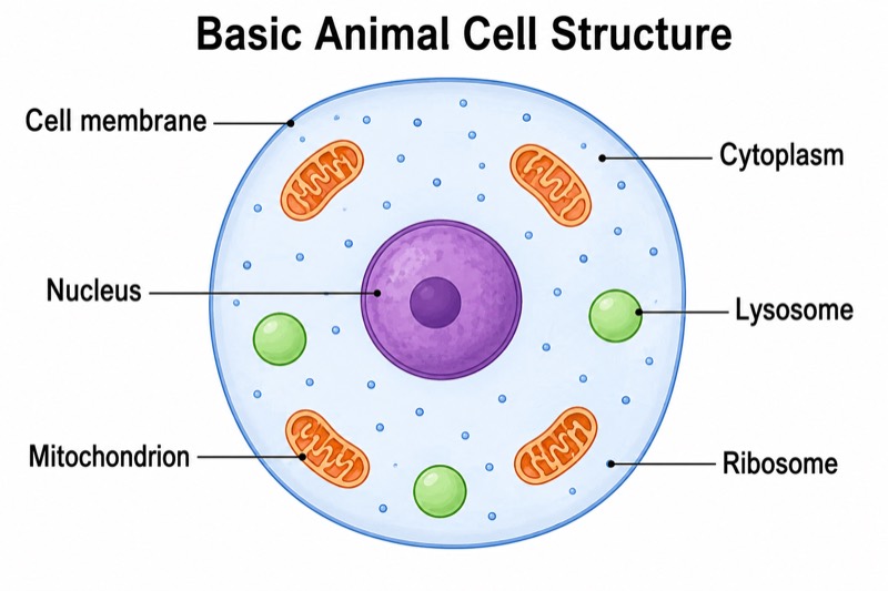
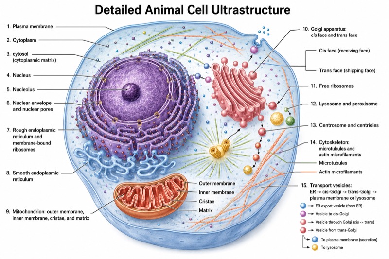
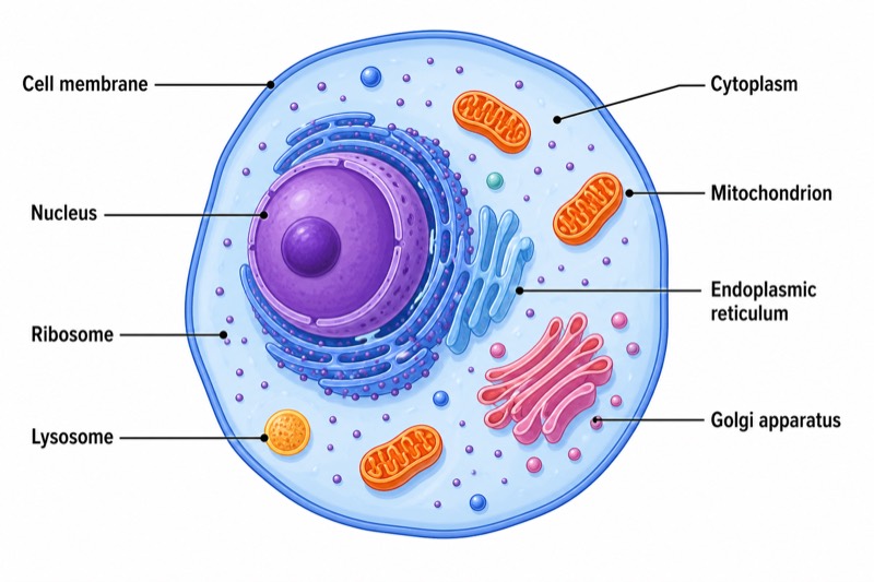
An animal cell structure diagram shows the main parts of a typical animal cell and their functions. The cell membrane forms the flexible outer boundary and controls what enters and leaves. Inside, the cytoplasm surrounds the organelles and is the site of many chemical reactions. The nucleus contains genetic material and directs cell activities. Mitochondria release usable energy through aerobic respiration, while ribosomes build proteins. The diagram should also show the endoplasmic reticulum, Golgi apparatus, and lysosomes in distinct colors and with clear labels.
The organelles work together as an organized system. Instructions in the nucleus are used by ribosomes to assemble proteins. Some ribosomes attach to the rough endoplasmic reticulum, where newly made proteins are folded and transported. Vesicles carry them to the Golgi apparatus, which modifies, sorts, and packages them for use or secretion. Mitochondria provide energy for these activities. Lysosomes contain enzymes that break down waste and worn-out cell parts. Meanwhile, the cell membrane regulates exchange with the environment, and the cytoplasm provides the fluid medium in which organelles and reactions are supported.
What a correct diagram must include
- Cell membrane — draw a thin, flexible outer boundary because animal cells do not have a cell wall.
- Cytoplasm — fill the space inside the cell membrane with a light color and place the organelles within it.
- Nucleus — draw a large, usually rounded structure containing genetic material and label it as the control center of the cell.
- Mitochondria — show several bean-shaped organelles with folded inner membranes to represent the site of aerobic respiration.
- Endoplasmic reticulum — draw a folded membrane network near the nucleus; add dots to rough endoplasmic reticulum to represent attached ribosomes.
- Golgi apparatus — use a stack of curved, flattened membrane sacs, often with small vesicles budding from the edges.
- Ribosomes — represent these as tiny dots, either free in the cytoplasm or attached to rough endoplasmic reticulum.
- Lysosomes — draw small, round membrane-bound sacs and identify them as organelles containing digestive enzymes.
Common mistakes
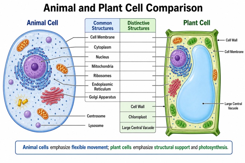
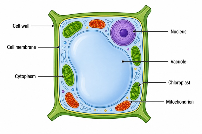
- Adding a cell wall or chloroplasts — these are typical plant cell structures and should not appear in a standard animal cell diagram.
- Drawing the cell as a rigid rectangle — animal cells have a flexible cell membrane and are usually shown with a rounded or irregular outline.
- Confusing mitochondria with the Golgi apparatus — mitochondria have folded inner membranes, while the Golgi apparatus is a stack of curved sacs.
- Drawing ribosomes too large — ribosomes should appear as very small dots, not as major membrane-bound organelles.
- Placing organelles outside the cytoplasm or allowing label lines to cross — all organelles should lie inside the cell membrane, with clear label lines.
Teaching tips
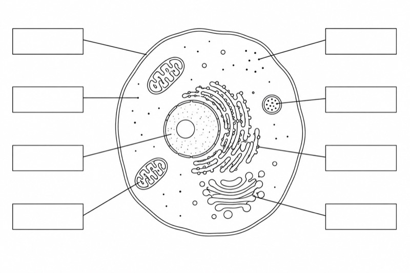
Use the diagram after introducing cells as the basic units of life. Ask students to identify each organelle, describe its function, and explain how its shape supports that function. Follow with relationship questions such as, “How does a protein move from a ribosome to the Golgi apparatus?” Students can then complete an unlabeled diagram or compare animal and plant cells. This supports common assessment points: matching structures to functions, distinguishing cell types, and tracing the roles of the nucleus, ribosomes, endoplasmic reticulum, Golgi apparatus, and mitochondria.
FAQ about this diagram
What controls the activities of an animal cell?
The nucleus controls many cell activities because it contains DNA, the genetic instructions for making proteins. These proteins influence cell structure, chemical reactions, growth, and repair.
How do ribosomes, the endoplasmic reticulum, and the Golgi apparatus work together?
Ribosomes assemble proteins, and attached ribosomes pass many new proteins into the rough endoplasmic reticulum. The proteins are then transported to the Golgi apparatus, where they are modified, sorted, and packaged.
What is the difference between the cell membrane and cytoplasm?
The cell membrane is the thin boundary that controls movement into and out of the cell. The cytoplasm is the jelly-like material inside the membrane where organelles are suspended and many chemical reactions occur.