Simple Animal Cell Diagram for Middle School
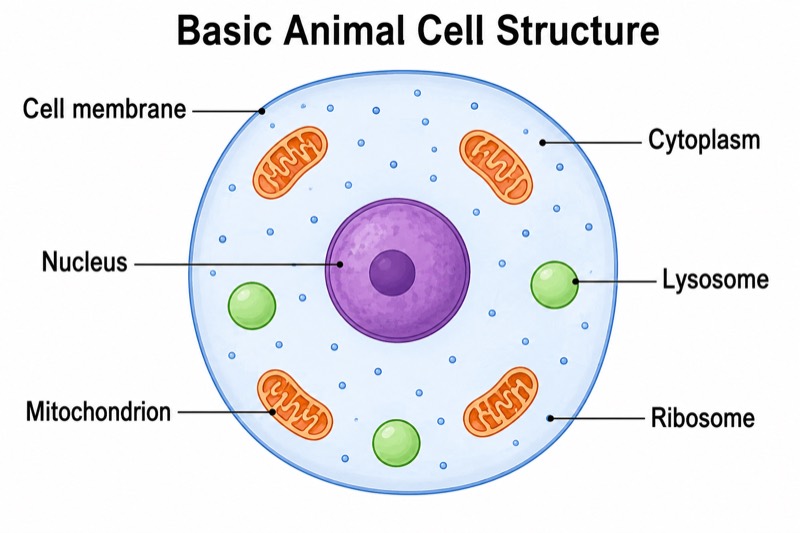
Simple Animal Cell Diagram for Middle School — an alternative version of the Animal Cell Structure Diagram, generated by AI and fully editable. Download the PNG for quizzes, homework or slides.
Labels included in this diagram
- Cell membrane
- Cytoplasm
- Nucleus
- Mitochondrion
- Ribosome
- Lysosome
✓ Accurate labels ✓ Edit text after generation ✓ PNG for papers, posters & slides
 OUTPUT · 16:9 · PNG
What this diagram shows
This simple animal cell diagram for middle school shows only six essential structures: the cell membrane, cytoplasm, nucleus, mitochondria, ribosomes, and lysosomes. Unlike the standard main diagram, it omits smaller organelles, internal membrane systems, and fine structural details. Rounded shapes, bright colors, large labels, balanced spacing, and generous white space make each part easy to identify. This version is useful when students are first learning cell organization because it reduces visual overload and keeps attention on the structures most often introduced in upper elementary and middle school science.
Read the diagram from the outside inward: the cell membrane forms the boundary, the cytoplasm fills the interior, and the nucleus is the main control center. Then locate the mitochondria, ribosomes, and lysosomes within the cytoplasm. Use label-to-structure matching before discussing each function. Compared with the standard main diagram, this simplified version is better for recognition, recall, and introductory assessment, but not for studying advanced organelle relationships. Teachers can also remove or cover the labels and ask students to identify each structure or add one short function beside every name.
Teaching tips
- In-class quiz: hide the six labels, number the structures, and ask students to write each name and one basic function within five minutes.
- Homework: have students redraw the simplified cell, color-code the six structures, and explain how the cell membrane, cytoplasm, and nucleus differ.
- Presentation slide: display the uncluttered diagram while introducing one structure at a time; use the standard main diagram later when adding more organelles and detail.
- Review outline or printed handout: place the diagram beside a six-row table where students record each structure's location and main function.
- Paper or report illustration: use it for a basic overview of animal cell organization, label it as simplified and not to scale, and choose the standard diagram if the discussion includes additional organelles.
FAQ about this diagram
Why does this version include only six cell structures?
The limited set matches the learning needs of beginners and prevents less important details from competing for attention. The standard main diagram is more appropriate after students can confidently identify these six structures.
Is this simplified diagram scientifically complete?
No. It is an instructional model that highlights the cell membrane, cytoplasm, nucleus, mitochondria, ribosomes, and lysosomes while intentionally omitting other organelles and microscopic details. Use it for introductory learning and review, not for advanced analysis of cell ultrastructure.