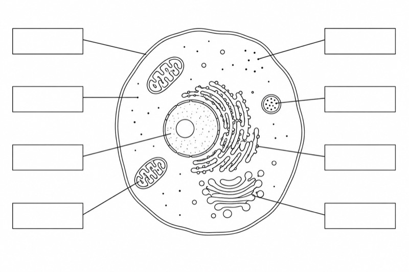
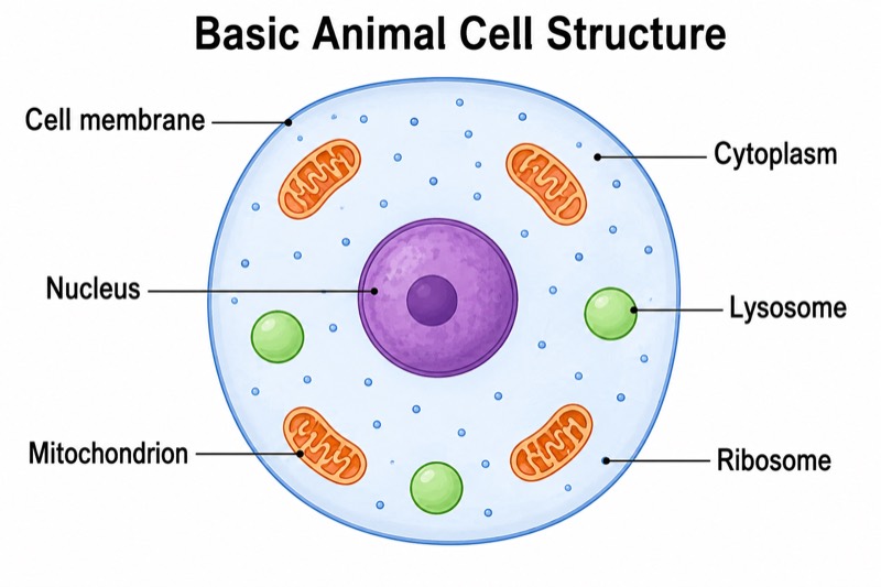
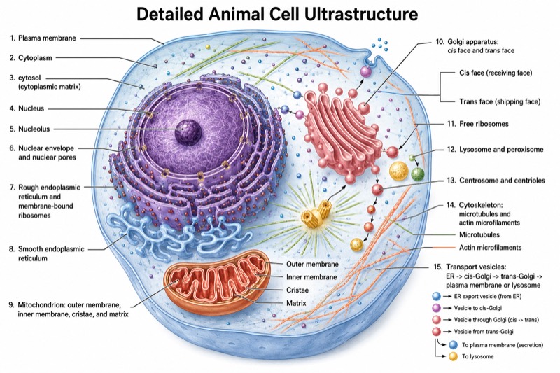
Animal and Plant Cell Comparison Diagram
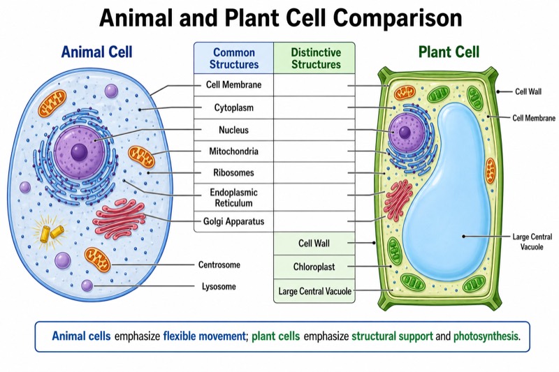
Animal and Plant Cell Comparison Diagram — an alternative version of the Animal Cell Structure Diagram, generated by AI and fully editable. Download the PNG for quizzes, homework or slides.
Labels included in this diagram
- Animal Cell
- Plant Cell
- Cell Membrane
- Cytoplasm
- Nucleus
- Mitochondria
- Ribosomes
- Endoplasmic Reticulum
- Golgi Apparatus
- Centrosome
- Lysosome
- Cell Wall
- Chloroplast
- Large Central Vacuole
✓ Accurate labels ✓ Edit text after generation ✓ PNG for papers, posters & slides
 OUTPUT · 16:9 · PNG
What this diagram shows
This Animal and Plant Cell Comparison Diagram places a rounded animal cell beside a rectangular plant cell, with corresponding organelles labeled across both illustrations. Unlike the standard animal cell structure diagram, which focuses on identifying parts within one animal cell, this version emphasizes similarities and differences between two eukaryotic cell types. A central comparison section lists shared structures—cell membrane, cytoplasm, nucleus, mitochondria, ribosomes, endoplasmic reticulum, and Golgi apparatus—and unique structures. Centrosomes and lysosomes are highlighted for the animal cell; the cell wall, chloroplasts, and large central vacuole are highlighted for the plant cell.
Read the diagram horizontally to match structures that occur in both cells, then use the contrasting colors and central text to identify cell-type-specific features. The bottom note connects structural differences to function, including photosynthesis, storage, support, intracellular digestion, and cell division. Use this comparison version when learners must explain relationships rather than merely name animal-cell organelles. It is especially useful after studying the standard main diagram: students can transfer prior knowledge, add plant-specific structures, and practice evidence-based comparisons such as why plant cells maintain a more regular shape or why chloroplasts are absent from animal cells.
Teaching tips
- In-class quiz: remove selected labels and ask students to identify each organelle, classify it as shared or cell-specific, and state one relevant function.
- Homework assignment: have students write a short comparison using the diagram, including at least three shared structures and two unique structures for each cell type.
- Presentation slide: display the side-by-side cells after teaching the standard animal cell diagram, then trace corresponding organelles across the page before discussing unique adaptations.
- Revision outline or printable handout: use the central lists as a compact checklist, and ask students to annotate the bottom functional differences in their own words.
- Essay or paper illustration: include the diagram when a written explanation compares eukaryotic cell organization, ensuring that the caption distinguishes typical textbook features from structures found in every individual cell.
FAQ about this diagram
Why use this comparison diagram instead of a standard animal cell structure diagram?
The standard diagram is better for learning the location and appearance of organelles within an animal cell. This comparison version is needed when the learning goal is to distinguish shared eukaryotic structures from animal-specific and plant-specific adaptations.
Are all listed structures unique to every animal or plant cell?
The diagram presents common textbook patterns, not absolute rules for every specialized cell. For example, some mature plant cells lack chloroplasts, and lysosome-like digestive compartments can vary; students should interpret the labels as typical comparative features.