Detailed Animal Cell Diagram (University Level)
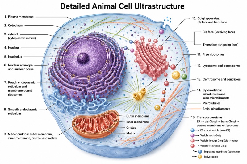
Detailed Animal Cell Diagram (University Level) — an alternative version of the Animal Cell Structure Diagram, generated by AI and fully editable. Download the PNG for quizzes, homework or slides.
Labels included in this diagram
- Plasma membrane
- Cytoplasm; cytosol (cytoplasmic matrix)
- Nucleus
- Nucleolus
- Nuclear envelope and nuclear pores
- Mitochondrion: outer membrane, inner membrane, cristae, and matrix
- Rough endoplasmic reticulum and membrane-bound ribosomes
- Smooth endoplasmic reticulum
- Golgi apparatus: cis face and trans face
- Free ribosomes
- Lysosome and peroxisome
- Centrosome and centrioles
- Cytoskeleton: microtubules and actin microfilaments
- Transport vesicles: ER -> cis-Golgi -> trans-Golgi -> plasma membrane or lysosome
✓ Accurate labels ✓ Edit text after generation ✓ PNG for papers, posters & slides
 OUTPUT · 16:9 · PNG
What this diagram shows
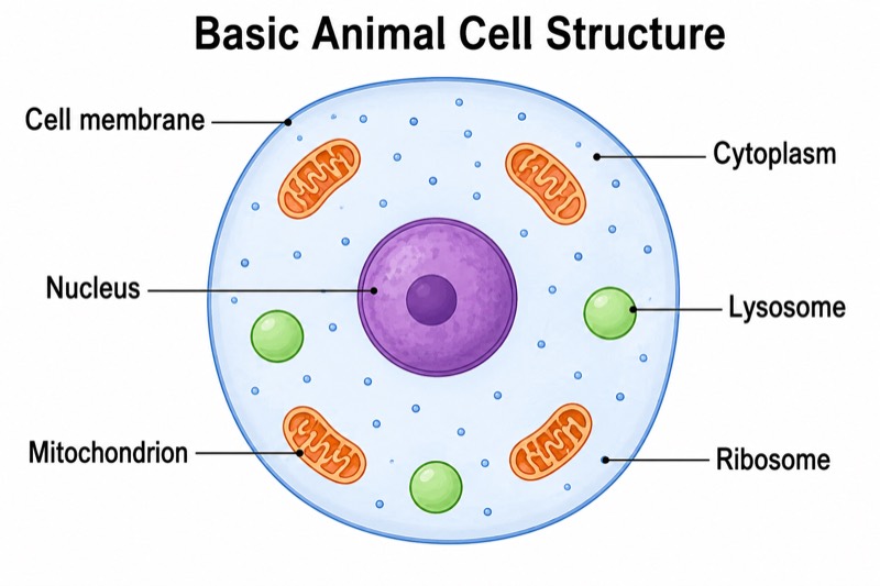
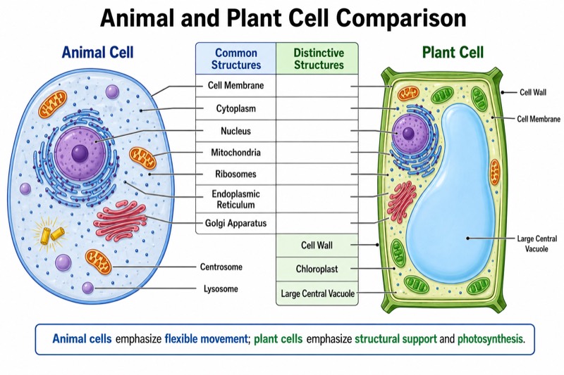
This detailed animal cell diagram (university level) expands the standard main diagram from a basic organelle overview into a research-oriented structural map. In addition to the plasma membrane, cytoplasm, nucleus, mitochondria, endoplasmic reticulum, Golgi apparatus, ribosomes, and lysosomes, it identifies the nucleolus, nuclear envelope and pores, rough and smooth endoplasmic reticulum, peroxisomes, centrosome, centrioles, cytosol, microtubules, and actin microfilaments. The Golgi cis and trans faces are distinguished, while a cutaway mitochondrion reveals cristae and matrix. This version is appropriate when subcellular organization matters more than introductory simplicity.
Read the figure by following spatial relationships and directional pathways rather than treating it as a list of isolated parts. Trace protein secretion from ribosomes on the rough endoplasmic reticulum through transport vesicles to the cis Golgi face, across Golgi cisternae, and out through the trans face. Compare that route with lipid synthesis in the smooth endoplasmic reticulum and intracellular digestion in lysosomes. Use the nuclear pores to connect nuclear and cytoplasmic processes, and examine how microtubules and microfilaments support organelle positioning and vesicle movement. Unlike the standard diagram, this version supports explanations of mechanisms, compartmentalization, and membrane trafficking.
Teaching tips
- In-class quizzes: remove selected labels and ask students to identify at least 12 structures, then add a second task requiring them to distinguish related features such as cytosol versus cytoplasm, or rough versus smooth endoplasmic reticulum.
- Homework and revision outlines: have students annotate each organelle with one structural feature and one function, then trace the secretory pathway from ribosome to plasma membrane. The extra detail makes this version more suitable than the standard main diagram for linking morphology to cellular processes.
- Lecture slides: reveal labels in stages—first major organelles, then nuclear structures, membrane-system subdivisions, cytoskeletal elements, and vesicle routes. This reduces visual overload while preserving the university-level relationships needed for discussions of protein sorting and intracellular transport.
- Printed handouts: provide one fully labeled reference copy and one blank or partially labeled copy. Students can color-code the endomembrane system, cytoskeleton, degradative compartments, and energy-related structures while checking that cis-to-trans Golgi polarity is correctly understood.
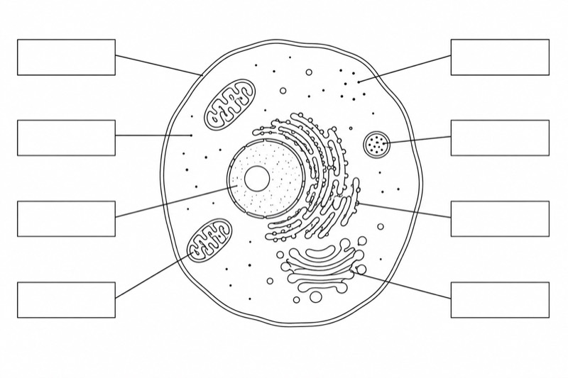
- Reports and academic figures: use the diagram as a conceptual overview when describing organelle interactions, vesicular trafficking, mitochondrial internal structure, or cytoskeletal organization. Because it is schematic rather than a microscopy image, the caption should state that organelle sizes, numbers, and distances are not drawn to scale.
FAQ about this diagram
Why use this detailed version instead of a standard animal cell diagram?
A standard diagram is best for first learning the major organelles, but it usually omits substructures and functional relationships. This university-level version distinguishes features such as nuclear pores, mitochondrial cristae, Golgi polarity, cytoskeletal fibers, and vesicle pathways, making it more useful for explaining mechanisms rather than simple identification.
Are all labeled structures visible in every real animal cell?
The structures are representative of a generalized eukaryotic animal cell, but their abundance, shape, and arrangement vary with cell type and physiological state. For example, secretory cells often contain extensive rough endoplasmic reticulum and Golgi stacks, whereas cells specialized for steroid synthesis tend to have more smooth endoplasmic reticulum.