Structure of a Nephron Generator
Free online Structure of a Nephron generator: get a fully labeled figure in about 90 seconds. The AI plans the must-have label list first, then renders a clean textbook-style diagram — every label editable afterwards, ready for papers, assignments and slides.
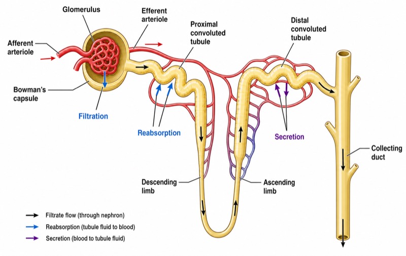
Labels included in this diagram
- Glomerulus
- Bowman's capsule
- Proximal convoluted tubule
- Descending limb
- Ascending limb
- Distal convoluted tubule
- Collecting duct
- Afferent arteriole
- Efferent arteriole
- Filtration
- Reabsorption
- Secretion
✓ Accurate labels ✓ Edit text after generation ✓ PNG for papers, posters & slides
 OUTPUT · 16:9 · PNG
What this diagram shows
A nephron structure diagram represents the kidney’s microscopic functional unit and the pathway by which blood plasma is converted into urine. It should show the renal corpuscle, formed by the glomerulus enclosed within Bowman’s capsule, followed by the proximal convoluted tubule, descending and ascending limbs of the loop of Henle, distal convoluted tubule, and collecting duct. The afferent arteriole supplies the glomerular capillaries, whereas the efferent arteriole carries blood away and gives rise to peritubular capillaries or the vasa recta. The diagram should also distinguish the renal cortex from the medulla and indicate the direction of tubular fluid flow.
The diagram links nephron anatomy to three major renal processes. Glomerular filtration occurs across the filtration barrier from glomerular capillary blood into Bowman’s space. Tubular reabsorption transfers water and useful solutes from the tubular lumen into the interstitial fluid and then the peritubular capillaries; it is especially extensive in the proximal tubule. Tubular secretion moves selected substances from blood or tubular cells into the lumen, mainly in the proximal and distal tubules. The descending limb is highly permeable to water, whereas the thick ascending limb reabsorbs ions but is effectively impermeable to water. Distal segments and collecting ducts provide hormonally regulated adjustment of electrolyte, acid–base, and water balance.
What a correct diagram must include
- Renal corpuscle: Draw a capillary tuft labeled glomerulus inside the cup-shaped Bowman’s capsule, leaving Bowman’s space between them.
- Afferent and efferent arterioles: Show the afferent arteriole entering and the efferent arteriole leaving the glomerulus; the afferent vessel is typically drawn wider to support filtration pressure.
- Tubular sequence: Connect Bowman’s capsule to the proximal convoluted tubule, descending limb, ascending limb, distal convoluted tubule, and collecting duct in the correct order.
- Loop of Henle: Extend the descending and ascending limbs into the medulla as a hairpin loop, with the descending limb preceding the ascending limb.
- Cortex and medulla: Place the renal corpuscle and convoluted tubules in the cortex, while the loop of Henle and collecting duct extend into the medulla.
- Blood supply around the tubule: Continue the efferent arteriole into peritubular capillaries or the vasa recta to show where reabsorbed substances enter the blood and secreted substances originate.
- Process arrows: Mark filtration from the glomerulus into Bowman’s space, reabsorption from the tubular lumen toward blood, and secretion from blood toward the tubular lumen.
- Flow direction: Add arrows showing blood flow from afferent arteriole to glomerulus to efferent arteriole, and filtrate flow from Bowman’s capsule toward the collecting duct.
Common mistakes
- Reversing the afferent and efferent arterioles or drawing the efferent arteriole as if it drains directly into a vein rather than forming a second capillary network.
- Placing filtration along the renal tubule; filtration occurs only at the renal corpuscle, whereas the tubules perform reabsorption and secretion.
- Drawing the distal convoluted tubule before the loop of Henle or connecting the proximal tubule directly to the collecting duct.
- Assigning water reabsorption equally to both limbs of the loop; the descending limb is water-permeable, while the ascending limb reabsorbs salts and is essentially water-impermeable.
- Treating the collecting duct as part of a single nephron without qualification; it receives tubular fluid from multiple nephrons and is part of the collecting system.
Teaching tips
Use the diagram after introducing renal gross anatomy and before teaching clearance, concentration, and acid–base regulation. Ask students to trace one water molecule from glomerular blood to final urine, then identify where it could be filtered, reabsorbed, or retained. A second prompt can compare the consequences of constricting the afferent versus efferent arteriole. Link each tubular segment to permeability, major transport functions, and hormonal control. The diagram directly supports examination questions on flow sequence, cortical versus medullary location, the countercurrent mechanism, glomerular filtration, and the distinction between reabsorption and secretion.
FAQ about this diagram
Where do filtration, reabsorption, and secretion occur in the nephron?
Filtration occurs from glomerular capillaries into Bowman’s space. Reabsorption and secretion occur along the renal tubule, with the proximal tubule handling the largest fraction of bulk transport and distal segments providing regulated adjustment.
Why are the descending and ascending limbs of the loop of Henle functionally different?
The descending limb is highly permeable to water and allows tubular fluid to become concentrated. The ascending limb, especially its thick segment, reabsorbs Na+, K+, and Cl− while remaining effectively impermeable to water, helping establish the medullary osmotic gradient.
What is the difference between the glomerulus and Bowman’s capsule?
The glomerulus is a tuft of fenestrated capillaries supplied by the afferent arteriole. Bowman’s capsule surrounds the glomerulus and receives the ultrafiltrate in Bowman’s space before it enters the proximal convoluted tubule.