Medical Illustration (Eye Anatomy) Generator
Free online Medical Illustration (Eye Anatomy) generator: get a fully labeled figure in about 90 seconds. The AI plans the must-have label list first, then renders a clean textbook-style diagram — every label editable afterwards, ready for papers, assignments and slides.
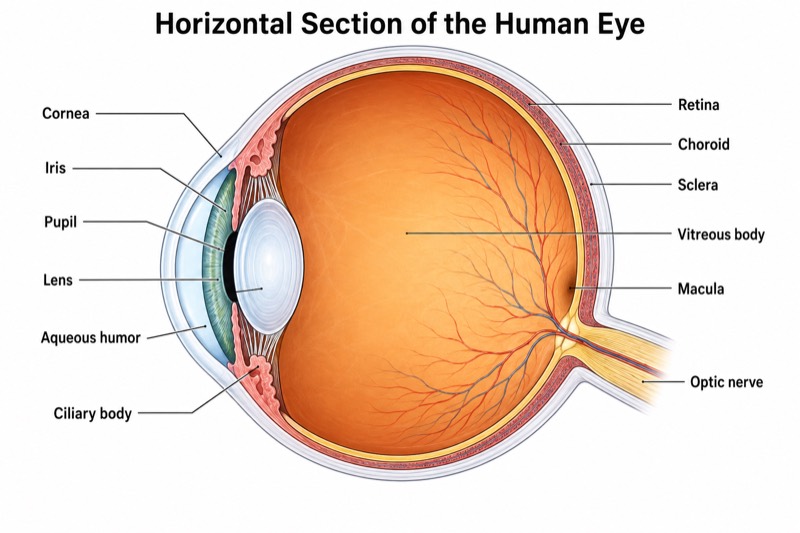
Labels included in this diagram
- Cornea
- Iris
- Pupil
- Lens
- Ciliary body
- Vitreous body
- Retina
- Choroid
- Sclera
- Macula
- Optic nerve
- Aqueous humor
✓ Accurate labels ✓ Edit text after generation ✓ PNG for papers, posters & slides
 OUTPUT · 16:9 · PNG
What this diagram shows
This medical illustration presents a horizontal section of the human eyeball, emphasizing the structures that transmit, focus, and detect light. Anteriorly, the transparent cornea and aqueous humor admit light, while the iris regulates the diameter of the pupil. The lens provides adjustable refractive power and is connected functionally to the ciliary body. The vitreous body occupies the large posterior cavity and supports the globe. The wall of the eye is shown in three principal layers: the inner retina, vascular choroid, and fibrous sclera. The macula is identified as the retinal region specialized for high-acuity central vision, and the optic nerve marks the exit of retinal ganglion cell axons.
The diagram should communicate the optical pathway and the anatomical relationships among ocular compartments. Light passes through the cornea, aqueous humor, pupil, lens, and vitreous body before reaching the retina. Corneal curvature supplies most of the eye's fixed refractive power, whereas contraction of the ciliary body changes lens shape during accommodation. The iris lies anterior to the lens and controls retinal illumination by altering pupil size. The retina converts focused light into neural signals; these signals travel through retinal circuitry and then enter the optic nerve. Outside the retina, the choroid provides vascular support and absorbs scattered light, while the sclera forms the strong outer coat that maintains shape and protects internal structures.
What a correct diagram must include
- Sectional orientation and proportions: Draw the eye as a near-spherical globe with a convex anterior cornea and a narrower optic nerve emerging posteriorly.
- Cornea, aqueous humor, iris, and pupil: Show the transparent cornea in front of the aqueous-filled anterior region, with the pupil represented as an opening in the iris rather than as a solid structure.
- Lens and ciliary body: Place the biconvex lens directly behind the pupil and connect its equatorial region to the ciliary body through the zonular apparatus when sufficient detail is required.
- Vitreous body: Depict it as the transparent gel filling most of the globe behind the lens, without confusing it with the smaller aqueous-filled spaces anterior to the lens.
- Retina, choroid, and sclera: Arrange these coats from inside to outside in the correct order—retina, choroid, then sclera—and use clearly distinguishable line weights or colors.
- Macula: Mark it on the posterior retina near the visual axis, lateral to the optic disc region, and avoid placing it directly where the optic nerve exits.
- Optic nerve: Draw it leaving the posterior globe and indicate continuity with retinal ganglion cell axons rather than portraying it as attached to the lens or vitreous body.
- Labeling system: Use straight, noncrossing leader lines, consistent English anatomical terms, and sufficient spacing so that labels do not obscure boundaries.
Common mistakes
- Drawing the pupil as a black anatomical disc; the pupil is an aperture in the iris through which light passes.
- Reversing the ocular coats; the correct sequence from the vitreous outward is retina, choroid, and sclera.
- Labeling the entire posterior retina as the macula or placing the macula at the optic nerve head; these are distinct retinal regions.
- Showing aqueous humor throughout the posterior segment; aqueous humor occupies the anterior and posterior chambers, whereas the vitreous body fills the vitreous chamber behind the lens.
- Placing the iris behind the lens or omitting its central opening; the iris lies anterior to the lens and forms the pupil.
Teaching tips
Use the illustration after introducing ocular anatomy and before teaching refraction, accommodation, or retinal physiology. Ask students first to trace the path of light from the cornea to the retina, then identify which structures refract light, regulate illumination, provide nutrition, or transmit neural signals. Follow with comparison questions such as aqueous humor versus vitreous body and macula versus optic nerve head. For assessment, remove selected labels, require the three ocular coats in correct order, and ask students to predict how corneal opacity, lens deformation, retinal damage, or optic nerve injury would alter vision.
FAQ about this diagram
Which structures refract light as it enters the eye?
The cornea provides most of the eye's refractive power because light crosses the air-cornea interface. The lens adds adjustable focusing power, especially during accommodation for near vision.
What is the difference between aqueous humor and the vitreous body?
Aqueous humor is a continuously produced fluid occupying the anterior and posterior chambers in front of the lens. The vitreous body is a transparent gel that fills the much larger vitreous chamber behind the lens and helps maintain globe shape.
How are the macula and optic nerve functionally different?
The macula is a specialized retinal region responsible for detailed central vision, with the foveal area providing the highest visual acuity. The optic nerve carries retinal ganglion cell axons toward the brain, and its exit point lacks photoreceptors.