Blood Circulation Diagram Generator
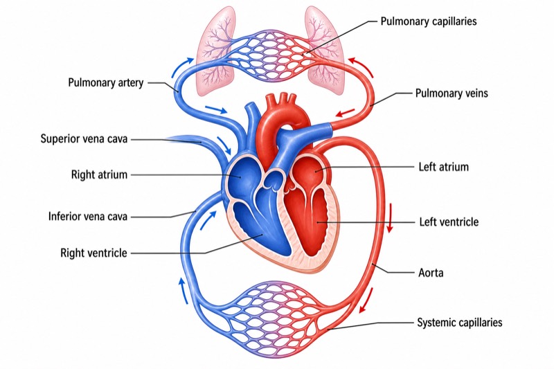
Free online Blood Circulation Diagram generator: get a fully labeled figure in about 90 seconds. The AI plans the must-have label list first, then renders a clean textbook-style diagram — every label editable afterwards, ready for papers, assignments and slides.
Labels included in this diagram
- Left ventricle
- Aorta
- Systemic capillaries
- Superior vena cava
- Inferior vena cava
- Right atrium
- Right ventricle
- Pulmonary artery
- Pulmonary capillaries
- Pulmonary veins
- Left atrium
✓ Accurate labels ✓ Edit text after generation ✓ PNG for papers, posters & slides
 OUTPUT · 16:9 · PNG
What this diagram shows
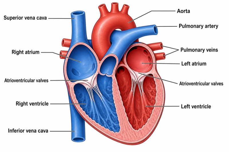
A human blood circulation diagram shows the double circulatory system formed by the systemic and pulmonary circuits. The heart functions as two coordinated pumps. The right side sends deoxygenated blood to the lungs, while the left side sends oxygenated blood to the body. Essential labels include the left ventricle, aorta, systemic capillaries, superior and inferior venae cavae, right atrium, right ventricle, pulmonary artery, pulmonary capillaries, pulmonary veins, and left atrium. Red should represent oxygenated blood and blue should represent deoxygenated blood, helping students compare blood oxygen levels along each pathway.
Blood flows in one continuous sequence: left ventricle → aorta → systemic capillaries → superior and inferior venae cavae → right atrium → right ventricle → pulmonary artery → pulmonary capillaries → pulmonary veins → left atrium → left ventricle. In systemic capillaries, oxygen and nutrients move from the blood into body tissues, while carbon dioxide enters the blood. In pulmonary capillaries, carbon dioxide diffuses into the alveoli and oxygen enters the blood. Arrows must show one-way flow, and the color should change only at the capillary exchange sites rather than inside the heart.
What a correct diagram must include
- Two clearly separated circuits: show the systemic circuit between the heart and body tissues and the pulmonary circuit between the heart and lungs.
- Four heart chambers: label the right atrium, right ventricle, left atrium, and left ventricle in their correct flow sequence.
- Major blood vessels: include the aorta, superior and inferior venae cavae, pulmonary artery, and pulmonary veins, connecting each to the correct chamber.
- Two capillary networks: draw systemic capillaries for tissue exchange and pulmonary capillaries for gas exchange in the lungs.
- Directional arrows: indicate uninterrupted one-way flow through vessels, chambers, and capillary beds.
- Accurate color coding: use red for oxygenated blood and blue for deoxygenated blood, changing color at the pulmonary and systemic capillaries.
- Complete circulation pathway: ensure that the route forms a closed loop from the left ventricle through both circuits and back to the left ventricle.
Common mistakes
- Reversing the pulmonary vessels: the pulmonary artery carries deoxygenated blood from the right ventricle to the lungs, while the pulmonary veins carry oxygenated blood to the left atrium.
- Defining arteries and veins by oxygen level: arteries carry blood away from the heart, whereas veins carry blood toward the heart.
- Connecting the venae cavae to the right ventricle instead of the right atrium, or connecting the pulmonary veins to the left ventricle instead of the left atrium.
- Changing red to blue inside a heart chamber; oxygen levels change mainly through gas exchange in systemic or pulmonary capillaries.
- Drawing both circuits as independent loops without showing that they operate in series as one continuous, closed circulation.
Teaching tips
Use the diagram after introducing heart structure and before explaining capillary exchange. Ask students to trace one red blood cell from the left ventricle until it returns there, naming every chamber, vessel, and capillary bed. Follow with questions such as, “Where does blood gain oxygen?” and “Why is the pulmonary artery blue?” The diagram directly supports assessment of circulation order, vessel classification, oxygenation changes, gas exchange, and the functional differences between systemic and pulmonary circulation.
FAQ about this diagram
What is the difference between systemic and pulmonary circulation?
Systemic circulation carries oxygenated blood from the left ventricle to body tissues and returns deoxygenated blood to the right atrium. Pulmonary circulation carries deoxygenated blood from the right ventricle to the lungs and returns oxygenated blood to the left atrium.
Why is the pulmonary artery shown in blue and the pulmonary veins in red?
Vessel names are based on the direction of blood flow relative to the heart, not oxygen content. The pulmonary artery carries deoxygenated blood away from the heart, while the pulmonary veins carry oxygenated blood toward the heart.
Where should the blood color change in the diagram?
Blue changes to red in the pulmonary capillaries as blood gains oxygen and releases carbon dioxide. Red changes to blue in the systemic capillaries as tissues receive oxygen and add carbon dioxide to the blood.