Anatomical Drawing (Muscular System) Generator
Free online Anatomical Drawing (Muscular System) generator: get a fully labeled figure in about 90 seconds. The AI plans the must-have label list first, then renders a clean textbook-style diagram — every label editable afterwards, ready for papers, assignments and slides.
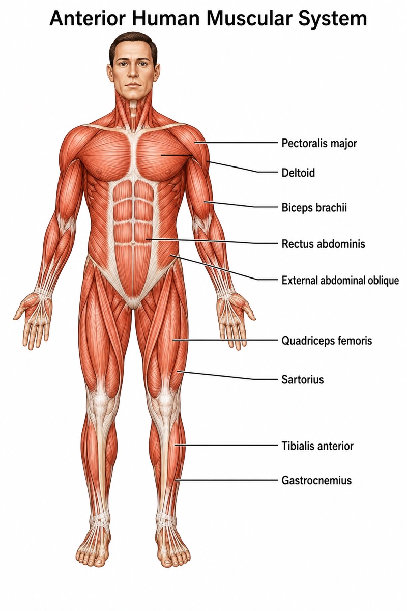
Labels included in this diagram
- Pectoralis major
- Deltoid
- Biceps brachii
- Rectus abdominis
- External abdominal oblique
- Quadriceps femoris
- Sartorius
- Tibialis anterior
- Gastrocnemius
✓ Accurate labels ✓ Edit text after generation ✓ PNG for papers, posters & slides
 OUTPUT · 16:9 · PNG
What this diagram shows
An anterior view of the human muscular system shows the superficial skeletal muscles that shape the front of the trunk and limbs. Pectoralis major covers the upper chest, while the deltoid forms the rounded shoulder and biceps brachii occupies the anterior arm. Rectus abdominis extends vertically along the abdominal wall, bordered laterally by external abdominal oblique. In the lower limb, quadriceps femoris dominates the anterior thigh, sartorius crosses it obliquely, and tibialis anterior lies beside the tibial crest. The upper contours of gastrocnemius may be visible around the calf, although most of this muscle is posterior.
These muscles form coordinated functional groups across major joints. Pectoralis major and deltoid move the humerus, whereas biceps brachii flexes the elbow and assists forearm supination. Rectus abdominis flexes the trunk, and external abdominal oblique contributes to trunk rotation, lateral flexion, and abdominal compression. Quadriceps femoris extends the knee, while sartorius crosses both the hip and knee to assist hip flexion, abduction, lateral rotation, and knee flexion. Tibialis anterior dorsiflexes and inverts the foot; gastrocnemius plantarflexes the ankle and helps flex the knee. Their arrangement illustrates how muscle position relative to a joint predicts mechanical action.
What a correct diagram must include
- Use the standard anatomical position — draw the body upright, facing forward, with the upper limbs at the sides and palms facing anteriorly so directional relationships remain correct.
- Show bilateral symmetry — paired muscles such as pectoralis major, deltoid, biceps brachii, rectus abdominis, and quadriceps femoris should appear on both sides of the body.
- Differentiate muscle from tendon — render muscle bellies as broad, fiber-textured regions and tendons or aponeuroses as narrower, paler connective-tissue structures.
- Map the trunk muscles accurately — place pectoralis major across the upper chest, rectus abdominis near the midline, and external abdominal oblique lateral to the rectus sheath.
- Represent quadriceps femoris as a group — indicate rectus femoris centrally, vastus lateralis laterally, and vastus medialis medially rather than treating the quadriceps as one undivided muscle.
- Draw sartorius as an oblique strap — it should run from the anterior superior iliac spine across the thigh toward the medial proximal tibia.
- Place lower-leg muscles by compartment — tibialis anterior belongs immediately lateral to the tibial crest, whereas gastrocnemius forms the posterior calf and is only partly visible in a strict anterior view.
- Use clear leader lines and precise labels — each line should terminate on the intended muscle belly without crossing unnecessarily or pointing to adjacent fascia, bone, or another muscle.
Common mistakes
- Labeling quadriceps femoris as a single muscle without recognizing its four components: rectus femoris, vastus lateralis, vastus medialis, and the deeper vastus intermedius.
- Drawing sartorius vertically down the thigh; its defining feature is its long oblique course from the lateral pelvis to the medial tibia.
- Placing tibialis anterior on the posterior calf or directly over the tibial crest instead of in the anterior compartment just lateral to the tibia.
- Showing the entire gastrocnemius prominently in a strict anterior view; it is a posterior muscle, although its proximal medial and lateral contours may be visible.
- Confusing biceps brachii with brachialis or triceps brachii and assigning elbow extension to it; biceps brachii primarily produces elbow flexion and forearm supination.
Teaching tips
Use the diagram after students have learned anatomical position, directional terminology, and major bones. Begin with identification questions, then ask students to predict each muscle’s action from its position relative to the shoulder, elbow, trunk, hip, knee, or ankle. Follow with comparisons such as tibialis anterior versus gastrocnemius or rectus abdominis versus external abdominal oblique. For assessment, require students to label muscles, classify them by body region, state principal actions, and identify whether each muscle crosses one or more joints. The figure also supports clinical questions about gait, posture, muscle weakness, and peripheral nerve injury.
FAQ about this diagram
Why is quadriceps femoris considered a muscle group rather than one muscle?
Quadriceps femoris consists of rectus femoris, vastus lateralis, vastus medialis, and vastus intermedius. All converge into the quadriceps tendon and extend the knee, but only rectus femoris also crosses the hip and contributes to hip flexion.
How can muscle location be used to predict movement?
A muscle usually moves a joint according to the side on which its line of pull passes. For example, tibialis anterior passes anterior to the ankle axis and produces dorsiflexion, whereas gastrocnemius passes posterior to the axis and produces plantarflexion.
What is the functional difference between rectus abdominis and external abdominal oblique?
Rectus abdominis primarily flexes the trunk and stabilizes the pelvis. External abdominal oblique compresses the abdominal contents and contributes to trunk flexion, ipsilateral lateral flexion, and contralateral rotation.