AI Scientific Image Generator
Free online AI Scientific Image generator: get a fully labeled figure in about 90 seconds. The AI plans the must-have label list first, then renders a clean textbook-style diagram — every label editable afterwards, ready for papers, assignments and slides.
Labels included in this diagram
- Mouse Model
- Tissue Collection
- Tissue Dissociation
- Cell Culture
- Centrifugation
- Nucleic Acid Extraction
- PCR Amplification
- Sequencing
- Bioinformatics Analysis
✓ Accurate labels ✓ Edit text after generation ✓ PNG for papers, posters & slides
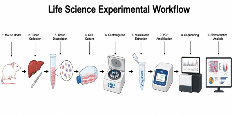 OUTPUT · 16:9 · PNG
What this diagram shows
This scientific illustration summarizes an experimental workflow for converting a biological question into interpretable molecular data. It begins with a Mouse Model representing the experimental system, followed by Tissue Collection from anatomically defined sites. Tissue Dissociation converts intact tissue into a cell suspension, while Cell Culture maintains or expands selected populations under controlled conditions. Centrifugation separates cells, debris, or liquid fractions according to sedimentation behavior. Nucleic Acid Extraction then isolates DNA or RNA suitable for downstream assays. PCR Amplification enriches defined target sequences, Sequencing determines nucleotide composition, and Bioinformatics Analysis transforms raw reads into quality-controlled, biologically meaningful results.
The components should form a left-to-right chain connected by directional arrows, with each transition showing a change in sample state. The mouse provides tissue; tissue is mechanically or enzymatically dissociated; and the resulting suspension may be cultured before centrifugation collects cells or separates fractions. The selected pellet or supernatant must be identified because this decision determines what enters Nucleic Acid Extraction. Extracted DNA can proceed directly to target-specific PCR, whereas extracted RNA normally requires reverse transcription before amplification. PCR products undergo quality control and sequencing preparation before Sequencing. Bioinformatics Analysis follows data generation and includes read quality assessment, trimming, alignment or assembly, quantification, statistical testing, and biological interpretation.
What a correct diagram must include
- Directional workflow — arrange all stages from Mouse Model to Bioinformatics Analysis on one horizontal axis and use consistent arrows to prevent ambiguity about sequence.
- Distinct sample states — visually differentiate whole tissue, dissociated cells, cultured cells, centrifuged fractions, purified nucleic acid, amplicons, and digital sequence reads.
- Mouse Model and Tissue Collection — indicate the experimental organism, treatment groups, anatomical sampling site, and collection point because these variables define biological context.
- Tissue Dissociation and Cell Culture — show tissue becoming a single-cell suspension and depict culture vessels only when cells are maintained, selected, stimulated, or expanded.
- Centrifugation outcome — draw a tube with a visible pellet and supernatant, clearly marking which fraction continues to the next step.
- Nucleic Acid Extraction and PCR Amplification — distinguish purified DNA or RNA from amplified targets and include reverse transcription when RNA is the starting material.
- Sequencing interface — show that validated amplicons or prepared libraries enter the sequencing stage rather than depicting untreated cells as direct sequencing input.
- Bioinformatics endpoint — represent raw reads, quality control, alignment or assembly, quantification, statistical analysis, and interpretable plots as connected computational outputs.
Common mistakes
- Placing Cell Culture as an obligatory step for every tissue experiment; freshly dissociated cells may instead proceed directly to sorting, centrifugation, or extraction.
- Drawing Centrifugation without identifying the retained pellet or supernatant, which obscures the biological material carried forward.
- Treating Tissue Dissociation and Nucleic Acid Extraction as equivalent; the former separates cells from tissue, whereas the latter releases and purifies DNA or RNA.
- Showing RNA entering conventional PCR directly; RNA workflows require reverse transcription to produce complementary DNA before amplification.
- Ending the workflow at Sequencing and omitting read quality control, normalization, statistical testing, and biological interpretation.
Teaching tips
Use the illustration after introducing experimental design and before students read a primary research methods section. Ask learners to trace the physical form of the sample at every arrow, identify where biological replicates and controls enter, and predict the consequence of retaining the wrong centrifugation fraction. The figure can support assessment of pre-analytical variables, DNA-versus-RNA workflows, PCR specificity, sequencing inputs, and the distinction between wet-laboratory and computational stages. For an advanced exercise, students can annotate quality-control checkpoints, potential batch effects, and metadata required for reproducibility.
FAQ about this diagram
Why is Tissue Dissociation performed before Cell Culture?
Dissociation releases cells from the extracellular matrix and disrupts tissue architecture, producing a suspension suitable for plating or enrichment. Enzymatic and mechanical conditions must be optimized because excessive treatment can reduce viability and alter gene expression.
What does Centrifugation separate in this workflow?
Centrifugation separates components according to size, density, and sedimentation rate. Cells commonly form a pellet while soluble molecules remain in the supernatant, but the fraction retained depends on the experimental objective.
How are PCR Amplification, Sequencing, and Bioinformatics Analysis different?
PCR selectively increases the abundance of defined nucleic acid targets, whereas sequencing determines the nucleotide order of prepared molecules. Bioinformatics converts instrument output into filtered reads, alignments or assemblies, quantitative measurements, statistical results, and biological conclusions.