Detailed Plant Cell Diagram (University Level)
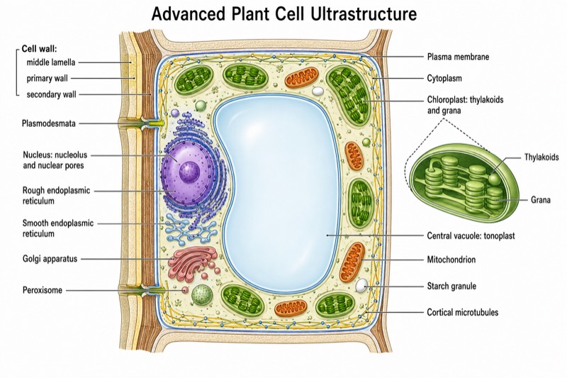
Detailed Plant Cell Diagram (University Level) — an alternative version of the Plant Cell Structure Diagram, generated by AI and fully editable. Download the PNG for quizzes, homework or slides.
Labels included in this diagram
- Cell wall: middle lamella, primary wall, secondary wall
- Plasma membrane
- Cytoplasm
- Plasmodesmata
- Nucleus: nucleolus and nuclear pores
- Chloroplast: thylakoids and grana
- Central vacuole: tonoplast
- Mitochondrion
- Rough endoplasmic reticulum
- Smooth endoplasmic reticulum
- Golgi apparatus
- Peroxisome
- Starch granule
- Cortical microtubules
✓ Accurate labels ✓ Edit text after generation ✓ PNG for papers, posters & slides
 OUTPUT · 16:9 · PNG
What this diagram shows
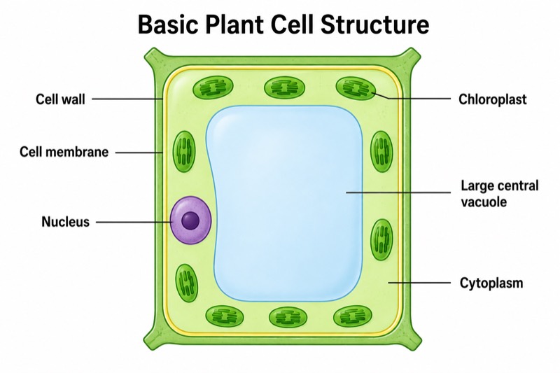
This detailed plant cell diagram (university level) extends the standard overview far beyond the cell wall, plasma membrane, cytoplasm, nucleus, chloroplasts, central vacuole, and mitochondria. It distinguishes the middle lamella, primary wall, secondary wall, plasmodesmata, tonoplast, nucleolus, nuclear pores, rough and smooth endoplasmic reticulum, Golgi apparatus, peroxisomes, starch grains, and microtubules. A cutaway chloroplast also reveals thylakoids and grana. Unlike the standard main diagram, which prioritizes rapid recognition of major organelles, this version shows the membranes, substructures, and cellular connections needed for university-level analysis.
Read the figure from the outer boundary inward: compare the middle lamella and wall layers, locate plasmodesmata crossing adjacent walls, and then distinguish the plasma membrane from the tonoplast surrounding the vacuole. Follow the non-crossing leader lines to connect each structure with its compartment and function. Use the enlarged chloroplast section to relate grana to photosynthetic reactions, and trace the rough ER, smooth ER, and Golgi apparatus as an endomembrane pathway. Choose this version when the standard main diagram lacks enough detail for explaining transport, secretion, wall development, organelle ultrastructure, or cytoskeletal organization.
Teaching tips
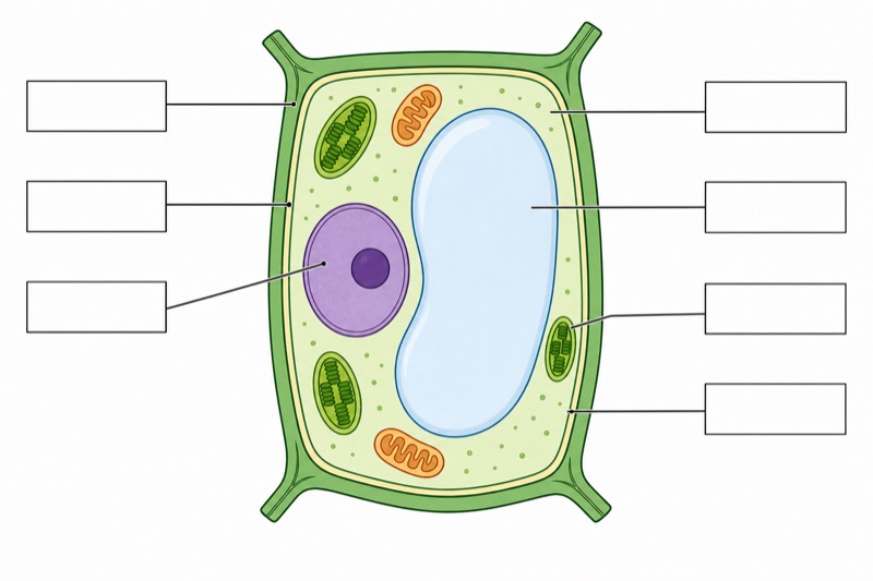
- In-class quiz: remove selected labels and ask students to identify at least 12 structures, while distinguishing commonly confused boundaries such as the cell wall, plasma membrane, and tonoplast.
- Homework: require students to annotate each labeled structure with one function and one structural relationship, such as nuclear pores controlling exchange or plasmodesmata connecting neighboring cells.
- Lecture slides: reveal the labels in stages, beginning with the standard major organelles and then adding wall layers, endomembrane components, peroxisomes, microtubules, and chloroplast ultrastructure.
- Revision outline or printed handout: pair the diagram with a table comparing location, membrane number, internal organization, and function; use the detailed version because the standard main figure cannot support these comparisons.
- Research report or thesis background figure: use the academically detailed layout to orient readers before discussing cell-wall deposition, symplastic transport, vacuolar compartmentation, photosynthesis, or organelle interactions; verify that every label matches the terminology used in the text.
FAQ about this diagram
Why use this university-level version instead of a standard plant cell diagram?
A standard diagram is best for learning the major organelles quickly, but it usually merges wall layers and omits membrane systems, pores, cytoskeletal elements, and organelle substructures. This detailed version is necessary when students must explain mechanisms such as secretion, symplastic transport, secondary-wall formation, or thylakoid organization.
Which distinctions are most important when studying this diagram?
Do not treat the cell wall, plasma membrane, and tonoplast as a single boundary: they have different locations and functions. Also distinguish primary wall from secondary wall, rough ER from smooth ER, and the chloroplast envelope from internal thylakoids arranged into grana.